INTRODUCTION
Epidermoid cysts rarely develop in the cecum, yet they have been reported in other internal organs, including the testes, spleen, accessory spleen, liver and kidneys [1]. Only eight cases of cecal epidermoid cysts have been reported in the English literature [1, 2, 3, 4, 5, 6, 7, 8]. Epidermoid cysts are generally considered to be sequestration cysts that may be congenital or acquired. An acquired epidermoid cyst develops in a patient with a history of abdominal trauma or surgery whereas a congenital epidermoid cyst may be the result of an aberrant embryogenic ectodermal implantation during embryogenesis. We report the case of a cecal epidermoid cyst in an elderly woman with no history of abdominal surgery or trauma. The cyst was discovered incidentally and was resected by using an ileocecectomy.
CASE REPORT
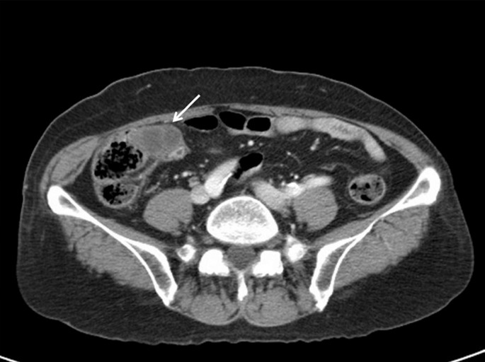
A 63-year-old female was admitted to Kyung Hee University Medical Center after an incidental finding of a right colonic mass during her regular health check-up. She underwent surgical excision of a submandibular gland due to a Wharton's duct stone. She was asymptomatic. On physical examination, there were no signs of fever or acute abdomen and no palpable abdominal mass. Colonoscopy revealed a mucosal tumor in the cecum without mucosal abnormalities. On abdominal computed tomography (CT), there was a 3.8-cm exophytic mass with internal low attenuation between the cecum and the terminal ileum (Fig. 1). The radiographic impression was that this mass was a gastrointestinal stromal tumor (GIST) and likely was not an adenocarcinoma. On further exploration, a round, soft, exophytic mass was identified at the cecal wall, 2.0 cm from the appendix orifice. After surgical investigation of the mass, an ileocecectomy was performed.
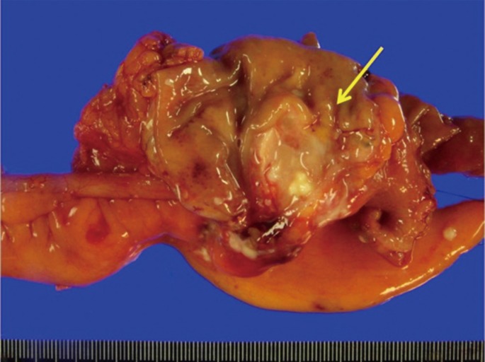
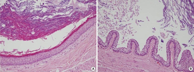
Macroscopically, the surface of the mass revealed an irregular cystic lesion filled with a yellowish-gray, cheesy material (Fig. 2). No connection between the cyst and the cecal lumen was observed. The mucosal and serosal surfaces of the cecum were unremarkable. Histologically, the wall of the cyst was surrounded in its entirety by a cecal muscularis propria. Cells lining the cyst formed a mature, keratinized, and stratified squamous epithelium with a granular layer (Fig. 3A). The focal area of cystic wall showed nonkeratinizing squamous epithelium without a granular layer (Fig. 3B). The lumen of the cyst was filled with mature, dense keratin. Skin adnexa were completely absent. No other ectodermal, endodermal or mesenchymal elements were noted. The pathological diagnosis was an epidermoid cyst.
DISCUSSION
Epidermoid cysts are generally believed to be sequestration cysts that may be congenital or acquired. Congenital epidermoid cysts are related to implantation of ectodermal elements at the time of closure of the neural groove or to the coalescence of other epithelial fusion lines, which typically occur in the head, neck, and anorectal areas and frequently involve the central nervous system and the intraspinal regions [9]. Epidermoid cysts can occur in the testes, spleen, accessory spleen, liver, and kidneys [1]. Acquired epidermoid cysts are traumatic or iatrogenic and may be caused by the implantation of the epidermis in locations favorable for growth [4].
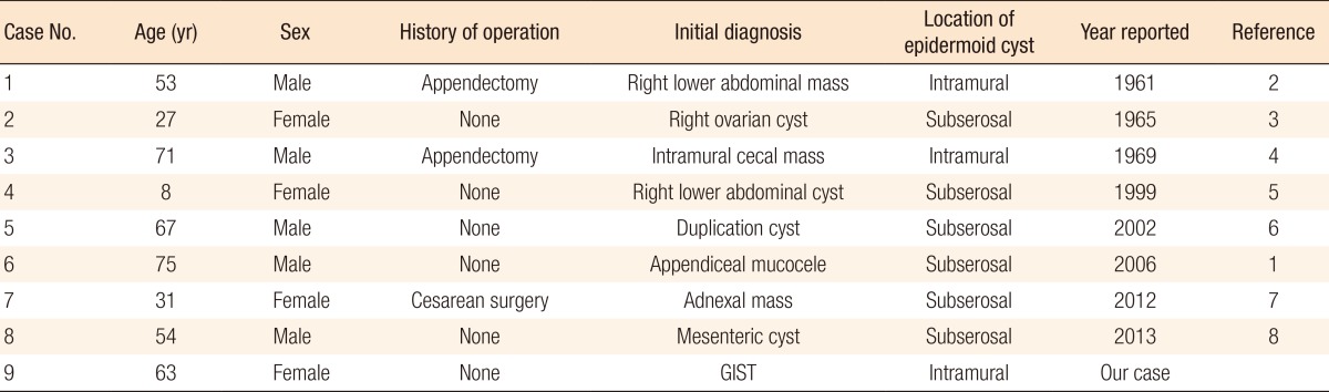
Epidermoid cysts of the cecum are extremely rare, with only eight reported cecal epidermoid cysts in the English literature (Table 1) [1, 2, 3, 4, 5, 6, 7, 8]. Three of the reported cecal epidermoid cyst cases were associated with a history of abdominal surgery. Two had undergone an appendectomy 12 years and 16 years prior to the diagnosis of a cecal epidermoid cyst [2, 4]. One patient had had a cesarean section [7]. These cases were attributed to iatrogenic implantation of epidermal fragments via a scalpel, needle or clamp during the operation [2, 4, 7]. The remaining patients had no history of abdominal surgery, and their cecal epidermoid cysts were considered to be congenital lesions. Andiran et al. [5] suggested that an inclusion or closure line of an epidermal structure may occur when the cecum re-enters the abdominal cavity during intrauterine rotation in the final steps of gut development. This encasing of an epidermal structure may result in later development of a cecal epidermoid cyst. Our patient had no history of abdominal surgery; therefore, her epidermoid cyst may have occurred due to an aberrant, embryonic, ectodermal implantation during embryogenesis.
The differential diagnosis of an epidermoid cyst is rarely considered in patients with cecal cystic lesions. Because epidermoid cysts of the cecum are mostly located in the subserosal area [1, 3, 5, 6, 7, 8], On CT imaging, these cysts may be confused with other intra-abdominal cystic lesions, including mesenteric cysts, lymphatic cysts, appendiceal mucoceles or duplication cysts [1, 8]. Especially, in female patients, they may initially be diagnosed as a right adnexal mass or cyst, so the differential diagnosis of an ovarian cyst or a cecal epidermoid cyst must be considered [3, 7]. Interestingly, our patient had an initial diagnosis of a GIST based on CT findings. This initial diagnosis was likely based on the unusual intramuscular location of the epidermoid cyst. In a previous case of a 71-year-old male patient, an epidermoid cyst within the cecal wall was considered to be an intramural cecal mass based on radiologic findings; the mass was then resected locally [4]. Grossly and histologically, epidermoid cysts resemble dermoid cysts. Dermoid cysts are lined with mature, squamous, epithelial cells and specialized skin structures, such as hair follicles, sweat glands and sebaceous glands. However, epidermoid cysts lack specialized skin structures, which differentiates them from dermoid cysts [9].
In conclusion, the development of an epidermoid cyst in the cecum, especially in the intramuscular region, is a rare occurrence; thus, epidermoid cysts are very rarely included in the differential diagnosis of cecal masses. From this case, we found that, given their location on radiographic imaging, cecal epidermoid cysts can be confused with mesenchymal cecal masses such as GISTs. Therefore, the possibility of an epidermoid cyst should be considered in the differential diagnosis for submucosal or intramuscular masses in the cecal area.