Warning: fopen(/home/virtual/colon/journal/upload/ip_log/ip_log_2025-05.txt): failed to open stream: Permission denied in /home/virtual/lib/view_data.php on line 95 Warning: fwrite() expects parameter 1 to be resource, boolean given in /home/virtual/lib/view_data.php on line 96 Primary Extrauterine Endometrial Stromal Sarcoma in the Sigmoid Colon
Skip Navigation
Skip to contents

Ann Coloproctol : Annals of Coloproctology

OPEN ACCESS
SEARCH
Search

Articles

Page Path
HOME > Ann Coloproctol > Volume 31(2); 2015 > Article
Case Report
Primary Extrauterine Endometrial Stromal Sarcoma in the Sigmoid Colon
Hyun-Jin Son, Joo-Heon Kim, Dong-Wook Kang, Hye-Kyung Lee, Mee-Ja Park, Seung Yun Lee
Annals of Coloproctology 2015;31(2):68-73.
DOI: https://doi.org/10.3393/ac.2015.31.2.68
Published online: April 30, 2015

Department of Pathology, Eulji University School of Medicine, Daejeon, Korea.

Correspondence to: Hyun-Jin Son, M.D. Department of Pathology, Eulji University Hospital, 95 Dunsanseo-ro, Seo-gu, Daejeon 302-799, Korea. Tel: +82-42-611-3451, Fax: +82-42-611-3459, shjpathol@eulji.ac.kr
• Received: December 11, 2014   • Accepted: February 6, 2015

© 2015 The Korean Society of Coloproctology

This is an open-access article distributed under the terms of the Creative Commons Attribution Non-Commercial License (http://creativecommons.org/licenses/by-nc/3.0/) which permits unrestricted non-commercial use, distribution, and reproduction in any medium, provided the original work is properly cited.

prev next
  • 6,583 Views
  • 65 Download
  • 8 Web of Science
  • 11 Crossref
  • 7 Scopus
  • An endometrial stromal sarcoma (ESS) is an uncommon uterine neoplasm, and its primary occurrence in the intestine as an extrauterine ESS (EESS) is exceedingly rare. We hereby report a primary EESS arising in the sigmoid colon with a review of the literature. A 52-year-old woman presented with bloody stool and underwent a colon fiberscopy, which revealed a fungating mass obstructing the lumen at the distal sigmoid. A laparoscopic low anterior resection was performed, and an umbilicated polypoid mass was identified; on section, it had infiltrated the mesocolic fat and measured 3.8 cm × 2.5 cm. The tumor showed geographic sheets or nests composed of relatively monotonous stromal cells, expansion or infiltration to the proper muscle and mesocolic fat, and extensive lymphovascular invasion and metastasis to regional lymph nodes and the pelvic peritoneum. The tumor cells were strongly and diffusely immunoreactive for CD10, but negative for c-kit, CD34, and Dog1. Two months later, a hysterectomy with a bilateral salpingo-oophorectomy was performed, and no evidence of an ESS was found in the uterus.
An endometrial stromal sarcoma (ESS), an uncommon uterine mesenchymal neoplasm, accounts for approximately 0.2% of all uterine malignancies [1]. Its occurrence at extrauterine sites is very rare, but it has been reported to occur at the ovary, bowel wall, abdomen, peritoneum, pelvis, and vagina [2]. Primary gastrointestinal involvement is rare [3,4,5,6,7,8,9,10,11,12,13]. We hereby present a case of a primary low-grade extrauterine ESS (EESS) in the sigmoid colon with a review of the literature. No evidence of a primary uterine ESS was found.
A 52-year-old woman was transferred from an outside clinic in order to evaluate the bloody stool and a pedunculated mass in the sigmoid colon that had been detected in a rigid sigmoid-scope examination. She was suffering from constipation, unspecified abdominal pain and hematochezia for a month. Her past and family histories were all unremarkable. Laboratory findings were unremarkable, except for mild anemia.
Colon fiberscopy was performed and revealed a fungating mass obstructing the lumen at the distal sigmoid colon (20 cm from the anal verge) (Fig. 1A). Under the impression of colon cancer, a biopsy was taken and suggested the possibility of a gastrointestinal stromal tumor (GIST) showing uncertain malignant potential. Abdominal computed tomography showed an eccentric and irregular wall thickening with enhancement measuring about 5 cm and perirectal lymph node enlargement (T3 or T4aN1M0, stage IIIB) (Fig. 1B). A laparoscopic low anterior resection was carried out, and an umbilicated polypoid mass measuring about 3.8 cm × 2.5 cm was identified in the distal sigmoid colon; on serial sections, it was found to have infiltrated the mesocolic fat (Fig. 1C, D). No dilation of the proximal bowel side to the mass was identified.
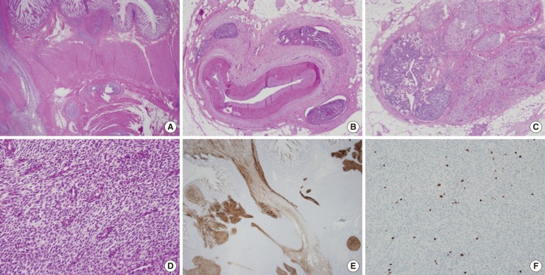
On histopathologic findings, the tumor showed geographic sheets or nests expanding to or infiltrating into the proper muscle and mesocolic fat (Fig. 2A), extensive lymphovascular invasion (Fig. 2B), and metastasis to regional lymph nodes (Fig. 2C) and to the pelvic peritoneum. In spite of extensive sampling, no focus of endometriosis was found in the resected colon specimen. Uniformly small, bland tumor cells with rounded-ovoid nuclei, evenly dispersed chromatin, no or inconspicuous nucleoli, and scanty cytoplasm with indistinct cell borders were noticed (Fig. 2D). Small, prominent arterioles were scattered among the stromal tumor cells. No mitosis or necrosis was identified. The tumor cells were strongly and diffusely immunoreactive for CD10 (1:50, Novocastra, Newcastle, UK) (Fig. 2E), estrogen receptor (ER; 1:40, Novocastra), and progesterone receptor (PR; 1:200, Novocastra), but negative for c-kit (1:600, Dako, Carpinteria, CA, USA), CD34 (predilution, Novocastra), Dog1 (1:500, Dako), α-smooth muscle actin (1:100, Novocastra), S100 protein (1:200, Novocastra), desmin (1:50, Dako) and pan-cytokeratin (1:200, Novocastra). The Ki-67 (1:100, Dako) labeling index was 8% (Fig. 2F).
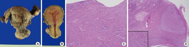
Endometrial curettage was conducted postoperatively and showed secretory hyperplasia and no evidence of ESS. Two months later, a total abdominal hysterectomy with a bilateral salpingo-oophorectomy was performed. The uterus measured 6.0 cm × 10.0 cm × 4.5 cm, and upon cross section, the uterine wall was thickened up to 3.2 cm and revealed an ill-defined trabeculated area with several hemorrhagic foci and a myomatous mass in the low uterine segment (Fig. 3A, B). Microscopically, it was diagnosed as adenomyosis, an intramural leiomyoma, and a relatively atrophic endometrium (Fig. 3C). Grossly, the bilateral adnexae were unremarkable, but microscopic foci of the endometrial stromal nodule were identified in the right and left ovaries and measured 0.6 cm and 0.8 cm, respectively (Fig. 3D). However, no focus of endometriosis was found in the uterus or bilateral adnexae. The patient was alive four months after the resection with no evidence of recurrence.
An ESS arising outside the uterus at extraovarian sites in the absence of a primary uterine lesion is extremely rare, and only several case reports are present in the English literature. The abdomen and the pelvis are the most common locations for an EESS, and its occurrence in the colonic wall is exceedingly rare, with only 9 reported cases in the literature (Table 1) [4,5,6,7,8,9,10,11]. Together with our case, 10 cases of an EESS in the colon have been reported, and the ages of the patients ranged from 38 to 80 years (mean, 52.2 years). The symptoms at presentation were abdominal pain, difficult defecation, hematochezia and alteration of bowel habits. The sigmoid colon and rectum are the most common sites of occurrence (9 out of 10 cases), which reflects the areas of the bowel having the highest incidence of endometriosis and the anatomic proximity to the ovary. Local dissemination was discovered at the time of the colonic resection in 8 out of the 10 cases, but no recurrence, except for one case, was found during the follow-up period (4 months to 4 years).
Associated foci of endometriosis in the colonic wall, peritoneal cavity or other sites were documented in 8 out of 9 previously reported cases. In our case, in spite of extensive sampling, no histological evidence of endometriosis was found in the specimens of the resected colon and both ovaries. Because the foci of endometriosis are almost always present in the vicinity of the EESS, one suggest that the primary EESS probably originated from an ectopic endometrial stroma of endometriosis, but our case implies that the mere absence of endometriosis does not preclude a primary EESS at that site. The pathogenesis of a primary EESS with no evidence of endometriosis is indefinite. The possibility is mentioned that the tumor might have originated from gland-poor foci of endometriosis [2], de novo from the peritoneal surface or the coelomic or subcoelomic multipotential epithelium [14, 15].
Malignant transformation is a rare, but well-known, complication of endometriosis, and most of these malignant neoplasms are endometrial adenocarcinomas (EAs). Yantiss et al. [11] reported 17 cases of gastrointestinal endometriosis complicated by neoplasms or precancerous changes. The neoplasms comprised 8 cases of EA, 4 cases of a Müllerian adenosarcoma, one case of an endometrioid adenofibroma of borderline malignancy with a carcinoma in situ, and one case of an EESS.
An unexpected location and an unusual presentation of an EESS may make the diagnosis challenging, despite classic histologic features. The GIST is the main differential diagnosis in the cases of an EESS arising in colon. The typical spiral arteriole-like vessels and perivascular whorl arrangement of monotonous spindle tumor cells should raise suspicion of an ESS. Immunohistochemistry is useful to distinguish these two entities. A GIST is known to be positively immunoreactive for c-kit, but an ESS is not. Moreover, an ESS shows diffuse immunoreactivity for CD10, ER, and PR. In our case, the tumor cells were strongly and diffusely immunoreactive for CD10, ER, and PR, but negative for c-kit.
In summary, we report a rare case of a primary EESS arising in the sigmoid colon in a 52-year-old woman with local dissemination to the pelvic peritoneum and both ovaries. Endometriosis was not found in specimens of the resected colon and both ovaries.

CONFLICT OF INTEREST: No potential conflict of interest relevant to this article was reported.

  • 1. Tavassoli FA, Devilee P. World Health Organization Classification of Tumors. Pathology and genetics of tumors of the breast and female genital organs. Lyon: IARC Press; 2003.
  • 2. Masand RP, Euscher ED, Deavers MT, Malpica A. Endometrioid stromal sarcoma: a clinicopathologic study of 63 cases. Am J Surg Pathol 2013;37:1635–1647. ArticlePubMed
  • 3. Jin M, Reynolds JP, Odronic SI, Wakely PE Jr. Primary gastric extra-uterine endometrial stromal sarcoma. Ann Diagn Pathol 2014;18:187–190. ArticlePubMed
  • 4. Ayuso A, Fadare O, Khabele D. A case of extrauterine endometrial stromal sarcoma in the colon diagnosed three decades after hysterectomy for benign disease. Case Rep Obstet Gynecol 2013;2013:202458.ArticlePubMedPMC
  • 5. Chen CW, Ou JJ, Wu CC, Hsiao CW, Cheng MF, Jao SW. High-grade endometrial stromal sarcoma arising from colon endometriosis. Int J Colorectal Dis 2007;22:1551–1553. ArticlePubMed
  • 6. Cho HY, Kim MK, Cho SJ, Bae JW, Kim I. Endometrial stromal sarcoma of the sigmoid colon arising in endometriosis: a case report with a review of literatures. J Korean Med Sci 2002;17:412–414. ArticlePubMedPMC
  • 7. Kovac D, Gasparovic I, Jasic M, Fuckar D, Dobi-Babic R, Haller H. Endometrial stromal sarcoma arising in extrauterine endometriosis: a case report. Eur J Gynaecol Oncol 2005;26:113–116. PubMed
  • 8. Baiocchi G, Kavanagh JJ, Wharton JT. Endometrioid stromal sarcomas arising from ovarian and extraovarian endometriosis: report of two cases and review of the literature. Gynecol Oncol 1990;36:147–151. ArticlePubMed
  • 9. Mourra N, Tiret E, Parc Y, de Saint-Maur P, Parc R, Flejou JF. Endometrial stromal sarcoma of the rectosigmoid colon arising in extragonadal endometriosis and revealed by portal vein thrombosis. Arch Pathol Lab Med 2001;125:1088–1090. ArticlePubMedPDF
  • 10. Bosincu L, Massarelli G, Cossu Rocca P, Isaac MA, Nogales FF. Rectal endometrial stromal sarcoma arising in endometriosis: report of a case. Dis Colon Rectum 2001;44:890–892. ArticlePubMed
  • 11. Yantiss RK, Clement PB, Young RH. Neoplastic and pre-neoplastic changes in gastrointestinal endometriosis: a study of 17 cases. Am J Surg Pathol 2000;24:513–524. ArticlePubMed
  • 12. Kim L, Choi SJ, Park IS, Han JY, Kim JM, Chu YC, et al. Endometrial stromal sarcoma of the small bowel. Ann Diagn Pathol 2008;12:128–133. ArticlePubMed
  • 13. Oliva E, Clement PB, Young RH. Epithelioid endometrial and endometrioid stromal tumors: a report of four cases emphasizing their distinction from epithelioid smooth muscle tumors and other oxyphilic uterine and extrauterine tumors. Int J Gynecol Pathol 2002;21:48–55. ArticlePubMed
  • 14. Norris HJ, Taylor HB. Mesenchymal tumors of the uterus. I. A clinical and pathological study of 53 endometrial stromal tumors. Cancer 1966;19:755–766. ArticlePubMed
  • 15. Lauchlan SC. The secondary Müllerian system. Obstet Gynecol Surv 1972;27:133–146. ArticlePubMed
Fig. 1

(A) Colon fiberscopy reveals a fungating mass obstructing the lumen at the distal sigmoid colon (20 cm from the anal verge). (B) Contrast-enhanced axial abdomen computed tomography shows an eccentric and irregular wall thickening with enhancement in the distal sigmoid colon measuring about 5 cm and a pericolic lymph node enlargement. (C) Grossly, an umbilicated polypoid mass measuring about 3.8 cm × 2.5 cm is identified in the distal sigmoid colon, and (D) on serial sections, it was found to have infiltrated to the mesocolic fat and to involve a regional lymph node.

ac-31-68-g001.jpg
Fig. 2

(A) The tumor shows geographic sheets or nests expanding to or infiltrating into the proper muscle and mesocolic fat (H&E, ×12.5). (B) Extensive lymphovascular invasion (H&E, ×40) and (C) metastasis to regional lymph nodes (H&E, ×40) are seen. (D) Uniformly small, bland tumor cells have rounded-ovoid nuclei, evenly dispersed chromatin, no or inconspicuous nucleoli, and scanty cytoplasm with indistinct cell borders. Small, prominent arterioles are scattered among the stromal tumor cells (H&E, ×200). (E) The tumor cells are strongly and diffusely immunoreactive for CD10 (×12.5). (F) The Ki-67 labeling index is 8% (×100).

ac-31-68-g002.jpg
Fig. 3

(A) The uterus measures 6.0 cm × 10.0 cm × 4.5 cm, and bilateral adnexae are grossly unremarkable. (B) Upon cross sectioning, the uterine wall was found to have thickened by 3.2 cm, and an ill-defined trabeculated area with several hemorrhagic foci was revealed. (C) Microscopically, the endometrium is relatively atrophic, and the uterine wall is thickened due to adenomyosis (H&E, ×40). (D) A microscopic focus of the endometrial stromal nodule is identified in the ovary (H&E, ×40) (Inset: high power view of the endometrial stromal nodule [H&E, ×200]).

ac-31-68-g003.jpg
Table 1

Clinical and pathological features of an EESS arising in the colon

ac-31-68-i001.jpg

EESS, extrauterine endometrial stromal sarcoma; TAH, total abdominal hysterectomy; BSO, bilateral salpingo-oophorectomy; NED, no evidence of disease; LSO, left salpingo-oophorectomy; RSO, right salpingo-oophorectomy; NA, not available.

Figure & Data

References

    Citations

    Citations to this article as recorded by  
    • Intestinal and Liver Involvement by Endometrial Stromal Sarcoma: Expanding the Differential Diagnosis of Mesenchymal Tumors Involving the GI Tract
      Yujie Zhang, Saman S. Karimi, Domenika Ortiz Requena, Armen Khararjian, Tom Z. Liang, Chen Mayer, Jacqueline Birkness-Gartman, Tatianna Larman, Elizabeth Anne Montgomery, Lysandra Voltaggio
      Human Pathology.2025; : 105784.     CrossRef
    • Primary Low-Grade Extrauterine Endometrial Stromal Sarcoma with Sex Cord Elements Involving the Omentum
      Chikkanaganna P. Manjula, V. Suguna Beluru, B. K. Raju, Naina Mary Simon
      Archives of Medicine and Health Sciences.2022; 10(1): 87.     CrossRef
    • Endometrial stromal sarcoma of the sigmoid colon: a case report and literature review
      Takuya Tajiri, Kosuke Mima, Kosuke Kanemitsu, Toru Takematsu, Keisuke Kosumi, Mitsuhiro Inoue, Takao Mizumoto, Tatsuo Kubota, Reiji Muto, Toshihiko Murayama, Nobutomo Miyanari, Hideo Baba
      International Cancer Conference Journal.2021; 10(4): 294.     CrossRef
    • CT, MRI, and FDG-PET imaging findings of low-grade extrauterine endometrial stromal sarcoma arising from the mesentery: A case report
      Satoshi Suzuki, Ryo Kurokawa, Tetsushi Tsuruga, Mayuyo Mori‑Uchino, Haruka Nishida, Tomoyasu Kato, Hiroyuki Abe, Tetsuo Ushiku, Shiori Amemiya, Akira Katayama, Osamu Abe
      Radiology Case Reports.2021; 16(9): 2774.     CrossRef
    • Isolated ureteral extrauterine endometrial stromal sarcoma: an unusual presentation
      Raghavendran Asokan, Leena Dennis Joseph, Arthi Mohanendran, Sriram Krishnamoorthy
      BMJ Case Reports.2021; 14(9): e245099.     CrossRef
    • Primary colonic extrauterine endometrial stromal sarcoma: A case and review of the literature
      Kuhali Kundu, Theresa Kuhn, Adrian Kohut, Charles Staley, Krisztina Hanley, Namita Khanna
      Gynecologic Oncology Reports.2020; 32: 100578.     CrossRef
    • Low-grade Endometrial Stromal Sarcoma Presenting as a Sigmoid Mass
      So Ra Ahn, Joo Hyun Lee
      The Korean Journal of Gastroenterology.2020; 76(6): 322.     CrossRef
    • Extra-uterine low grade endometrioid stromal sarcoma arising from ovarian endometriosis: a case report and review of the literature
      Boubacar Efared, Ibrahim S. Sidibé, Fatimazahra Erregad, Nawal Hammas, Laila Chbani, Hinde El Fatemi
      Gynecologic Oncology Research and Practice.2019;[Epub]     CrossRef
    • Multifocal low-grade endometrial stromal sarcoma arising from pre-existing endometriosis in a hysterectomised patient: a case report
      Saadet Alan, Ercan Yilmaz, Fahriye Tecellioglu, Ayse Nur Akatli, Ebru Inci Coskun, Hasan Gokce
      Journal of Obstetrics and Gynaecology.2019; 39(8): 1177.     CrossRef
    • Primary extragenital endometrial stromal sarcoma of the lung: first reported case and review of literature
      Lara Alessandrini, Francesco Sopracordevole, Giulio Bertola, Simona Scalone, Martina Urbani, Gianmaria Miolo, Tiziana Perin, Fabrizio Italia, Vincenzo Canzonieri
      Diagnostic Pathology.2017;[Epub]     CrossRef
    • An unexpected diagnosis of primary omental endometrial stromal sarcoma in a patient with acute right abdominal pain: A case report and review of literature
      Vered Buchholz, George Kiroff, Markus Trochsler, Harsh Kanhere
      International Journal of Surgery Case Reports.2017; 36: 8.     CrossRef

    • Cite this Article
      Cite this Article
      export Copy Download
      Close
      Download Citation
      Download a citation file in RIS format that can be imported by all major citation management software, including EndNote, ProCite, RefWorks, and Reference Manager.

      Format:
      • RIS — For EndNote, ProCite, RefWorks, and most other reference management software
      • BibTeX — For JabRef, BibDesk, and other BibTeX-specific software
      Include:
      • Citation for the content below
      Primary Extrauterine Endometrial Stromal Sarcoma in the Sigmoid Colon
      Ann Coloproctol. 2015;31(2):68-73.   Published online April 30, 2015
      Close
    • XML DownloadXML Download
    Figure
    • 0
    • 1
    • 2
    Primary Extrauterine Endometrial Stromal Sarcoma in the Sigmoid Colon
    Image Image Image
    Fig. 1 (A) Colon fiberscopy reveals a fungating mass obstructing the lumen at the distal sigmoid colon (20 cm from the anal verge). (B) Contrast-enhanced axial abdomen computed tomography shows an eccentric and irregular wall thickening with enhancement in the distal sigmoid colon measuring about 5 cm and a pericolic lymph node enlargement. (C) Grossly, an umbilicated polypoid mass measuring about 3.8 cm × 2.5 cm is identified in the distal sigmoid colon, and (D) on serial sections, it was found to have infiltrated to the mesocolic fat and to involve a regional lymph node.
    Fig. 2 (A) The tumor shows geographic sheets or nests expanding to or infiltrating into the proper muscle and mesocolic fat (H&E, ×12.5). (B) Extensive lymphovascular invasion (H&E, ×40) and (C) metastasis to regional lymph nodes (H&E, ×40) are seen. (D) Uniformly small, bland tumor cells have rounded-ovoid nuclei, evenly dispersed chromatin, no or inconspicuous nucleoli, and scanty cytoplasm with indistinct cell borders. Small, prominent arterioles are scattered among the stromal tumor cells (H&E, ×200). (E) The tumor cells are strongly and diffusely immunoreactive for CD10 (×12.5). (F) The Ki-67 labeling index is 8% (×100).
    Fig. 3 (A) The uterus measures 6.0 cm × 10.0 cm × 4.5 cm, and bilateral adnexae are grossly unremarkable. (B) Upon cross sectioning, the uterine wall was found to have thickened by 3.2 cm, and an ill-defined trabeculated area with several hemorrhagic foci was revealed. (C) Microscopically, the endometrium is relatively atrophic, and the uterine wall is thickened due to adenomyosis (H&E, ×40). (D) A microscopic focus of the endometrial stromal nodule is identified in the ovary (H&E, ×40) (Inset: high power view of the endometrial stromal nodule [H&E, ×200]).
    Primary Extrauterine Endometrial Stromal Sarcoma in the Sigmoid Colon

    Clinical and pathological features of an EESS arising in the colon

    EESS, extrauterine endometrial stromal sarcoma; TAH, total abdominal hysterectomy; BSO, bilateral salpingo-oophorectomy; NED, no evidence of disease; LSO, left salpingo-oophorectomy; RSO, right salpingo-oophorectomy; NA, not available.

    Table 1 Clinical and pathological features of an EESS arising in the colon

    EESS, extrauterine endometrial stromal sarcoma; TAH, total abdominal hysterectomy; BSO, bilateral salpingo-oophorectomy; NED, no evidence of disease; LSO, left salpingo-oophorectomy; RSO, right salpingo-oophorectomy; NA, not available.


    Ann Coloproctol : Annals of Coloproctology Twitter Facebook
    TOP