Warning: fopen(/home/virtual/colon/journal/upload/ip_log/ip_log_2025-05.txt): failed to open stream: Permission denied in /home/virtual/lib/view_data.php on line 95 Warning: fwrite() expects parameter 1 to be resource, boolean given in /home/virtual/lib/view_data.php on line 96 Colonic Metastasis Presenting as an Intraluminal Fungating Mass 8 Years After Surgery for Ovarian Cancer
Skip Navigation
Skip to contents

Ann Coloproctol : Annals of Coloproctology

OPEN ACCESS
SEARCH
Search

Articles

Page Path
HOME > Ann Coloproctol > Volume 31(5); 2015 > Article
Case Report
Colonic Metastasis Presenting as an Intraluminal Fungating Mass 8 Years After Surgery for Ovarian Cancer
Jeong Rye Kim, Bong Man Kim, You Me Kim, Won Ae Lee1, Hwan Namgung2
Annals of Coloproctology 2015;31(5):198-201.
DOI: https://doi.org/10.3393/ac.2015.31.5.198
Published online: October 31, 2015

Department of Radiology, Dankook University College of Medicine, Cheonan, Korea.

1Department of Pathology, Dankook University College of Medicine, Cheonan, Korea.

2Department of Surgery, Dankook University College of Medicine, Cheonan, Korea.

Correspondence to: Hwan Namgung, M.D. Departments of Surgery, Dankook University College of Medicine, 119 Dandae-ro, Dongnam-gu, Cheonan 31116, Korea. Tel: +82-41-550-3929, Fax: +82-41-556-3878, gsnamgung@dankook.ac.kr
• Received: December 24, 2014   • Accepted: April 16, 2015

© 2015 The Korean Society of Coloproctology

This is an open-access article distributed under the terms of the Creative Commons Attribution Non-Commercial License (http://creativecommons.org/licenses/by-nc/3.0/) which permits unrestricted non-commercial use, distribution, and reproduction in any medium, provided the original work is properly cited.

prev next
  • 5,640 Views
  • 43 Download
  • 4 Web of Science
  • 5 Crossref
  • 5 Scopus
  • We report a case of colonic metastasis from ovarian cancer presented as an intraluminal fungating mass mimicking primary colon cancer 8 years after surgery for ovarian cancer. A 70-year-old woman presented with constipation. She had undergone an extended total abdominal hysterectomy with bilateral salpingo-oophorectomy for an ovarian papillary serous cystadenocarcinoma 8 years earlier. Colonoscopy showed a large fungating mass 10 cm from the anal verge that was suspected to be colorectal cancer. A computed tomography scan showed a bulky intraluminal fungating mass in the rectosigmoid junction. After a lower anterior resection and a pathologic diagnosis, a diagnosis of a papillary serous adenocarcinoma due to metastasis from an ovarian tumor was made for this patient.
Epithelial ovarian carcinomas accounting for 85% to 90% of ovarian cancers show aggressive behavior [1]. Epithelial ovarian carcinomas tend to spread as direct invasion, and intraperitoneal seeding, and rarely as hematogenous metastasis [2]. If colonic metastasis from ovarian cancer presents as an intraluminal fungating mass, differentiating it from the primary colon cancer can be difficult. This report describes the case of a patient who had under-gone surgery for advanced ovarian cancer and had done well for 8 years, but then presented with a metastasis on the rectosigmoid junction as an intraluminal colonic mass.
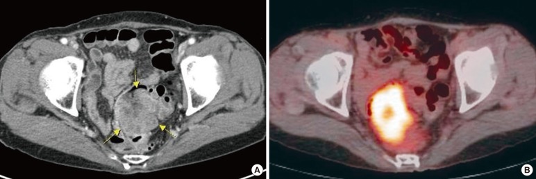
A 70-year-old woman presented with constipation in August 2013. Colonoscopy had been performed at an outside institution. A fungating mass of 6 cm in size was observed 10 cm from the anal verge and was thought to be colorectal cancer (Fig. 1). A biopsy was performed, and the pathologic diagnosis was a metastatic papillary serous adenocarcinoma. The patient's serum cancer antigen 125 (CA-125) was slightly elevated (63.2 U/mL; reference, 0-55 U/mL), but her serum carcinoembryonic antigen (CEA) was normal (1.5 ng/mL; reference, 0-7 ng/mL). The computed tomography (CT) scan showed a bulky intraluminal fungating mass involving the distal sigmoid colon and the proximal rectum, as well as several small lymph nodes in the superior rectal, left common iliac, and para-aortic nodal stations. Positron emission tomography-CT revealed a high fluoro-deoxyglucose uptake in the colonic mass and in the regional and the para-aortic lymph nodes, suggesting metastatic lymphadenopathy (Fig. 2).
The patient had undergone an extended total abdominal hysterectomy with a bilateral salpingo-oophorectomy for ovarian cancer in June 2005. Pathologic examination revealed a papillary serous cystadenocarcinoma in the right ovary. The tumor involved the contralateral ovary, the bilateral mesosalpinges, and the pelvic peritoneum, and according to the International Federation of Gy-necology and Obstetrics staging system, the tumor was stage IIIB. After surgery, the patient was treated with six cycles of paclitaxel, and for eight years, no evidence of recurrence or metastasis was observed. The patient's serum CA 125 level had been 24.6 U/mL before the first surgery and remained in the normal range until May 2012.
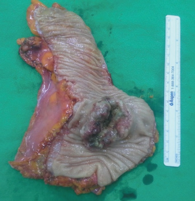
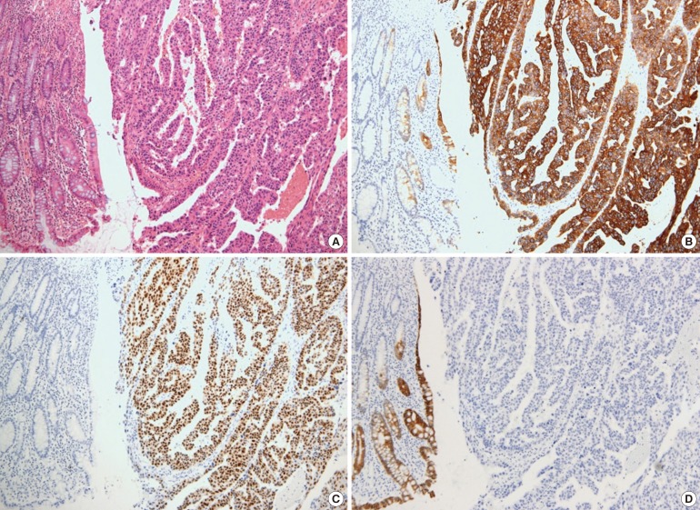
The patient underwent a lower anterior resection with lymphnode dissection for the regional and the para-aortic lymph nodes. A 6-cm intraluminal mass was noted in the rectosigmoid junction with no evidence of a peritoneal seeding nodule in the pelvic and the abdominal cavities. Macroscopically, the mucosa revealed a huge fungating tumor mimicking the primary colon cancer (Fig. 3). Microscopically, the tumor was an adenocarcinoma with papillary and glandular architectures (Fig. 4). The tumor had infiltrated through the mucosa to the subserosa. Histology data from the previous ovarian cancer were reviewed and were found to be identical to histology data for the present colon tumor. Immunohistochemically, both the ovarian and the colon cancers were diffusely positive for cytokeratin 7 (CK7) and estrogen receptor (ER), but negative for CK20 and mucin 2 (MUC2). The histological and the immunohistochemical consistency of the two tumors demonstrated that the colon tumor was a metastatic papillary serous adenocarcinoma from the ovary. Two out of forty-six lymph nodes had microscopic evidence of a metastatic carcinoma. The patient was treated with six cycles of docetaxel and carboplatin. At the time of this report, her serum CA 125 levels had been in the normal range for 15 months, and there had been no evidence of recurrent or metastatic disease.
Colonic metastases are classified by their mode of dissemination as follows: direct invasion from a contiguous or a noncontiguous primary tumor, intraperitoneal seeding, and embolic metastases [2]. Colonic metastasis from an ovarian epithelial neoplasm usually occurs by direct invasion or intraperitoneal seeding. Therefore, mostly the colonic serosa is initially affected, followed by bowel wall infiltration in an inward direction [34]. Colonic metastases from ovarian cancer can present as tethering, angulation, and extrinsic mass effects [5]. Zighelboim et al. [3] reported a case of recurrence of an epithelial ovarian carcinoma as an intraluminal bowel lesion with serosal sparing. It was considered to be the result of hematogenous dissemination of tumor cells with infiltration of the submucosal capillary network.
Recurrence of ovarian cancer as colonic metastasis after several years of being disease free is rare. Shibahara et al. [6] reported a case of one patient who underwent surgery to remove a papillary serous adenocarcinoma of both ovaries and then presented with colon metastasis 20 years later. Although the disease-free duration for the patient in the present case was shorter than that of the patient in the case described by Shibahara et al. [6], considering the aggressiveness of ovarian cancer, the emergence of metastasis after eight years is interesting. Kohyama et al. [7] reported another rare case of colon metastasis from ovarian cancer that presented with a protruding colonic mass with intussusception six years after surgery for ovarian cancer.
As in this case, if the colonic metastasis from ovarian cancer presents as an intraluminal fungating mass, a differential diagnosis of colonic metastasis and primary colon cancer based on the macroscopic appearance will be difficult. In such cases, serum CA 125 and CEA levels can be helpful for the differential diagnosis. Takahashi et al. [8] reported that in patients with a malignant ovarian tumor, 81.3% had positive serum CA 125. However, in patients with a serous cystadenocarcinoma, 100% showed positive serum CA 125.
Immunohistochemical studies for CK7 and CK20 can be very helpful to differentiate the origin of the tumor. In the study by Loy et al. [9], a CK7 positive/CK20 negative immunophenotype was 100% specific in differentiating a primary ovarian carcinoma from a metastatic colonic carcinoma. A CK7 negative/CK20 positive immunophenotype was 99% specific in differentiating a metastatic colonic carcinoma from a primary ovarian carcinoma [9]. Although these are less specific than CK immunostaining, positivity for CA 125, ER, and progesterone receptor immunostainings favor a primary ovarian carcinoma. In contrast, positivity for MUC2 and caudal-type homeobox 2 favors a primary colon cancer. In the present case, both ovarian and colon tumors were diffusely positive for CK7 and ER but completely negative for CK20 and MUC2, supporting both tumors being primary ovarian tumors.
In the past, a bowel resection in a sleeve fashion without a mesenteric resection has been shown to be an effective method of cytoreduction. However, O'Hanlan et al. [10] suggested that surgeons should consider a resection involving the bowel to have a 2- to 5-cm longitudinal negative margin with a wedge of mesentery, including the paracolic and intermediate level nodes, which is similar to the current standards for a resection of a primary bowel carcinoma. They recognized that ovarian cancer tended to spread longitudinally along the bowel wall's lymphatic channels into mesenteric nodes. After debulking surgery, a combination of a platinum compound (cisplatin or carboplatin) and taxane (paclitaxel or docetaxel) has been the standard approach for chemotherapy [1].
Distinguishing colonic metastasis from ovarian cancer and primary colon cancer may be difficult. In the case of a patient who presents with an intraluminal colonic mass with a history of ovarian cancer, having a high clinical suspicion of colonic metastasis from ovarian cancer is important. A biopsy followed by appropriate immunochemical staining is required for confirmation.

CONFLICT OF INTEREST: No potential conflict of interest relevant to this article was reported.

  • 1. American Cancer Society. Ovarian cancer [Internet]. Atlanta (GA): American Cancer Society; c2013.cited 2014 Jul 21. Available from: http://www.cancer.org/acs/groups/cid/documents/webcontent/003130-pdf.pdfPubMed
  • 2. Meyers MA, McSweeney J. Secondary neoplasms of the bowel. Radiology 1972;105:1–11. ArticlePubMed
  • 3. Zighelboim I, Broaddus R, Ramirez PT. Atypical sigmoid metastasis from a high-grade mixed adenocarcinoma of the ovary. Gynecol Oncol 2004;94:850–853. ArticlePubMed
  • 4. Reed E, Zerbe CS, Brawley OW, Bicher A, Steinberg SM. Analysis of autopsy evaluations of ovarian cancer patients treated at the National Cancer Institute, 1972-1988. Am J Clin Oncol 2000;23:107–116. ArticlePubMed
  • 5. Gore RM, Levine MS. Textbook of gastrointestinal radiology. 3rd ed. Philadelphia: Elsevier Saunders; 2008.PubMed
  • 6. Shibahara K, Endo K, Ikeda T, Sakata H, Sadanaga N, Morita M, et al. Colon metastasis 20 years after the removal of ovarian cancer: report of a case. Surg Today 2009;39:153–156. ArticlePubMed
  • 7. Kohyama M, Takesue Y, Ohge H, Sakashita M, Murakami Y, Sueda T. A case of colon metastasis from ovarian cancer presented with intussusception. J Jpn Surg Assoc 2005;66:2767–2771.Article
  • 8. Takahashi K, Shibukawa T, Moriyama M, Shirai T, Kijima S, Iwanari O, et al. Clinical usefulness and false-positive results of CA 125 as a tumor marker of ovarian cancer: a study on 674 patients. Jpn J Surg 1986;16:305–310. ArticlePubMed
  • 9. Loy TS, Calaluce RD, Keeney GL. Cytokeratin immunostaining in differentiating primary ovarian carcinoma from metastatic colonic adenocarcinoma. Mod Pathol 1996;9:1040–1044. PubMed
  • 10. O'Hanlan KA, Kargas S, Schreiber M, Burrs D, Mallipeddi P, Longacre T, et al. Ovarian carcinoma metastases to gastrointestinal tract appear to spread like colon carcinoma: implications for surgical resection. Gynecol Oncol 1995;59:200–206. ArticlePubMed
Fig. 1

Colonoscopy shows a large intraluminal fungating mass at the rectosigmoid junction of the colon about 10 cm from the anal verge.

ac-31-198-g001.jpg
Fig. 2

(A) The computed tomography (CT) image shows a heterogeneous enhanced mass in the rectosigmoid junction of colon. (B) The colonic mass shows a high fluoro-deoxyglucose uptake on the positron emission tomography-CT image.

ac-31-198-g002.jpg
Fig. 3

The colonic mucosa shows a huge fungating tumor simulating the primary colon cancer.

ac-31-198-g003.jpg
Fig. 4

Colon tumor. (A) Microscopically, the tumor is an adenocarcinoma with papillary and glandular architectures (H&E, ×100). The tumor cells are diffusely positive for cytokeratin 7 (CK7) (B) and estrogen receptor (C) and negative for CK20 (D) (immunohistochemistry, ×100).

ac-31-198-g004.jpg

Figure & Data

References

    Citations

    Citations to this article as recorded by  
    • A Case Report of Bilateral Endometrioid-Type Ovarian Carcinoma with Synchronous Dual Metastasis to the Colon
      W. T. N. Widanage, K. S. Nathawitharanalage, N. A. Kodithuwakku, A. A. S. Samarathunga, H. M. S. P. Rajaguru, K. P. Dissanayake, L. R. A. Wijesooriya, J. A. S. B. Jayasundara
      SN Comprehensive Clinical Medicine.2024;[Epub]     CrossRef
    • Metastasis of Ovarian Cancer to the Descending Colon
      Kentaro Abe, Hiroyuki Anzai, Satoko Eguchi, Masako Ikemura, Aya Shinozaki-Ushiku, Takahide Shinagawa, Hirofumi Sonoda, Yuichiro Yoshioka, Yuzo Nagai, Shinya Abe, Hiroyuki Matsuzaki, Yuichiro Yokoyama, Shigenobu Emoto, Koji Murono, Kazuhito Sasaki, Hiroaki
      Case Reports in Gastroenterology.2023; 17(1): 136.     CrossRef
    • Clinicopathologic Features of Gynecologic Malignancies Presenting Clinically as Colonic Malignancies
      Lanisha D Fuller, Andrew Dunn, Aaron R Huber, Monika Vyas, Raul S Gonzalez
      American Journal of Clinical Pathology.2022; 157(1): 82.     CrossRef
    • Rectorrhagia revealing colonic metastasis from an ovarian primary, an exceptional case report
      Rachid Jabi, Siham Elmir, Soumia El Arabi, Achraf Merry, Mohammed Bouziane
      International Journal of Surgery Case Reports.2021; 88: 106490.     CrossRef
    • Metastatic colon cancer of an ovarian cancer origin mimicking primary colon cancer: A case report
      Ji-Hyeon Park, Dong Hae Jung, Jeong-Heum Baek
      Korean Journal of Clinical Oncology.2018; 14(1): 53.     CrossRef

    • PubReader PubReader
    • Cite this Article
      Cite this Article
      export Copy Download
      Close
      Download Citation
      Download a citation file in RIS format that can be imported by all major citation management software, including EndNote, ProCite, RefWorks, and Reference Manager.

      Format:
      • RIS — For EndNote, ProCite, RefWorks, and most other reference management software
      • BibTeX — For JabRef, BibDesk, and other BibTeX-specific software
      Include:
      • Citation for the content below
      Colonic Metastasis Presenting as an Intraluminal Fungating Mass 8 Years After Surgery for Ovarian Cancer
      Ann Coloproctol. 2015;31(5):198-201.   Published online October 31, 2015
      Close
    • XML DownloadXML Download
    Figure
    • 0
    • 1
    • 2
    • 3
    Colonic Metastasis Presenting as an Intraluminal Fungating Mass 8 Years After Surgery for Ovarian Cancer
    Image Image Image Image
    Fig. 1 Colonoscopy shows a large intraluminal fungating mass at the rectosigmoid junction of the colon about 10 cm from the anal verge.
    Fig. 2 (A) The computed tomography (CT) image shows a heterogeneous enhanced mass in the rectosigmoid junction of colon. (B) The colonic mass shows a high fluoro-deoxyglucose uptake on the positron emission tomography-CT image.
    Fig. 3 The colonic mucosa shows a huge fungating tumor simulating the primary colon cancer.
    Fig. 4 Colon tumor. (A) Microscopically, the tumor is an adenocarcinoma with papillary and glandular architectures (H&E, ×100). The tumor cells are diffusely positive for cytokeratin 7 (CK7) (B) and estrogen receptor (C) and negative for CK20 (D) (immunohistochemistry, ×100).
    Colonic Metastasis Presenting as an Intraluminal Fungating Mass 8 Years After Surgery for Ovarian Cancer

    Ann Coloproctol : Annals of Coloproctology Twitter Facebook
    TOP