Warning: fopen(/home/virtual/colon/journal/upload/ip_log/ip_log_2025-06.txt): failed to open stream: Permission denied in /home/virtual/lib/view_data.php on line 95 Warning: fwrite() expects parameter 1 to be resource, boolean given in /home/virtual/lib/view_data.php on line 96 Single-center Experience of 24 Cases of Tailgut Cyst
Skip Navigation
Skip to contents

Ann Coloproctol : Annals of Coloproctology

OPEN ACCESS
SEARCH
Search

Articles

Page Path
HOME > Ann Coloproctol > Volume 35(5); 2019 > Article
Original Article
Single-center Experience of 24 Cases of Tailgut Cyst
Ahmad Sakr1,2, Ho Seung Kim1, Yoon Dae Han1, Min Soo Cho1, Hyuk Hur1, Byung Soh Min1, Kang Young Lee1, Nam Kyu Kim1orcid
Annals of Coloproctology 2019;35(5):268-274.
DOI: https://doi.org/10.3393/ac.2018.12.18
Published online: October 31, 2019

1Division of Colorectal Surgery, Department of Surgery, Severance Hospital, Yonsei University College of Medicine, Seoul, Korea

2Colorectal Surgery Unit, Department of General Surgery, Mansoura Faculty of Medicine, Mansoura University Hospitals, Mansoura, Egypt

Correspondence to: Nam Kyu Kim, M.D. Division of Colorectal Surgery, Department of Surgery, Yonsei University College of Medicine, 50-1 Yonsei-ro, Seodaemun-gu, Seoul 03722, Korea Tel: +82-2-2228-2100, Fax: +82-2-313-8289 E-mail: namkyuk@yuhs.ac
• Received: October 23, 2018   • Accepted: December 18, 2018

Copyright © 2019 The Korean Society of Coloproctology

This is an Open Access article distributed under the terms of the Creative Commons Attribution Non-Commercial License (http://creativecommons.org/licenses/by-nc/4.0/) which permits unrestricted non-commercial use, distribution, and reproduction in any medium, provided the original work is properly cited.

prev next
  • 10,833 Views
  • 288 Download
  • 34 Web of Science
  • 47 Crossref
  • 37 Scopus
See letter "Tailgut Cyst, Report of 24 Cases Single Center Experience" in Volume 35 on page 227.
  • Purpose
    Tailgut cysts are rare congenital or developmental lesions that arise from vestiges of the embryological hindgut. They are usually present in the presacral space. We report our single-center experience with managing tailgut cysts.
  • Methods
    We conducted a retrospective analysis of 24 patients with tailgut cyst treated surgically at the Colorectal Surgery Department of Severance Hospital, Yonsei University, Seoul, South Korea, between 2007–2018.
  • Results
    This study included 24 patients (18 females) with a median age of 51.5 years (range, 21–68 years). Ten cases were symptomatic and 14 were asymptomatic. Cysts were retrorectal in 21 patients. Cysts were below the coccyx level in 16 patients, opposite the coccyx in 6, and above the coccyx in 2. Cysts were supralevator in 5 patients, had a supra- and infralevator extension in 18 patients, and were infralevator in 1. Ten patients were managed using an anterior laparoscopic approach, 11 using a posterior approach, and 3 using a combined approach. Mean cyst size was 5.5 ± 2.7 cm. Postoperative complications were Clavien-Dindo (CD) classification grade II in 9 patients (37.5%) and CD grade III in 1 (4.2%). The posterior approach group showed the highest rate of complications (P = 0.021). Patients managed using a combined approach showed a larger cyst size (P < 0.001), longer operation times (P < 0.001), and a greater likelihood of tumor level above the coccyx (P = 0.002) compared to other approaches. The tumors of 2 male patients were malignant: 1 was a neuroendocrine tumor treated with radiotherapy, while the other was a closely followed adenocarcinoma. Median follow-up was 12 months (range, 1–66 months) with no recurrence.
  • Conclusion
    Tailgut cysts are uncommon but can cause perineal or pelvic pain. Complete surgical excision via an appropriate approach according to tumor size, location, and correlation with adjacent pelvic floor muscles is the key treatment.
Presacral tailgut cysts (retrorectal cystic hamartomas) are rare congenital lesions that are believed to arise from remnants of the hindgut when incomplete involution occurs during embryogenesis [1, 2]. These cysts are usually present in the presacral space, which is anteriorly bound by the rectum, posteriorly by the lower sacrum and coccyx, superiorly by the peritoneal reflection, inferiorly by the levator ani and pelvic floor, and laterally by the iliac vessels and ureters [3, 4]. However, cysts also occur in the prerectal, perianal, and perirenal regions [5-7]. These lesions are rare and can present diagnostic and management challenges. Most studies are case reports and case series [5, 8-12]. Surgical treatment is always required because of complications including malignant transformation, infection, and perianal fistula formation. The aim of this study was to report our single-center experience with tailgut cysts, including their management and surgical outcomes.
This retrospective case series included 24 patients with a pathologically confirmed tailgut cyst treated between January 2007 and August 2018 at the Colorectal Surgery Department of Severance Hospital, Yonsei University, Seoul, South Korea. The study protocol was approved by the Institutional Review Board of Yonsei University (approval number: 4-2018-0862). Written informed consents were waived off because of being retrospective study.
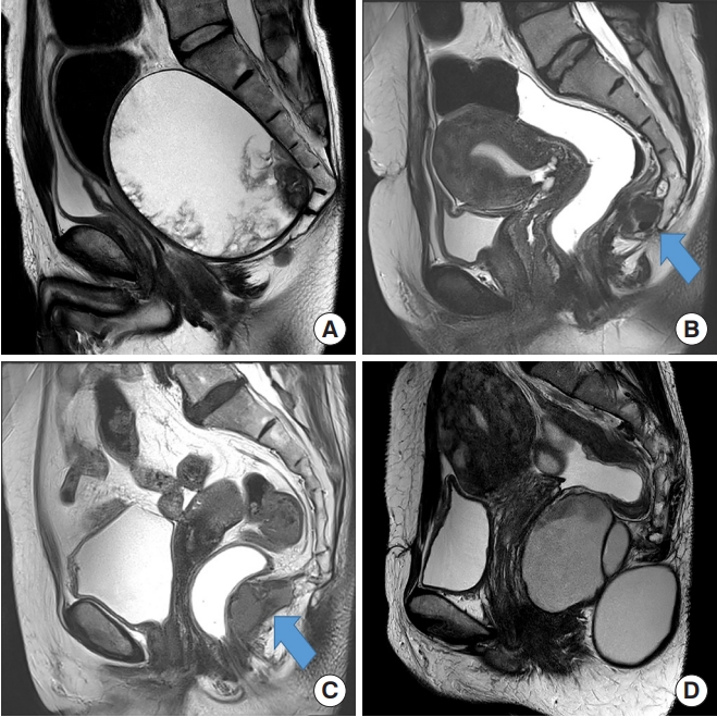
Patients’ medical records were reviewed for demographic variables, history, clinical presentation, previous surgical history, diagnostic methods, operative details, complications, histopathology, follow-up, and recurrence rates. All patients were diagnosed using pelvic magnetic resonance imaging (MRI) (Fig. 1).
Anterior (transabdominal), posterior (paracoccygeal), and combined (anterior + posterior) approaches were used. Most of the tumors were below the coccygeal level, so the posterior approach was usually used. However, the anterior approach was sometimes sufficient for removing tumors at the coccygeal level. For prerectal cysts, the anterior approach was used.
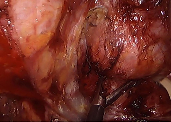
For the anterior approach, laparoscopy was used for all patients to enable complete mobilization of the rectum through the medial, lateral, posterior, deep posterior, and anterior dissection until the cyst was separated from the rectum for retrorectal cysts. For prerectal cysts in female patients, the aim was to mobilize the cyst by separating the rectum from the vagina, then completely excising the cyst via a small mini-laparotomy (Fig. 2).
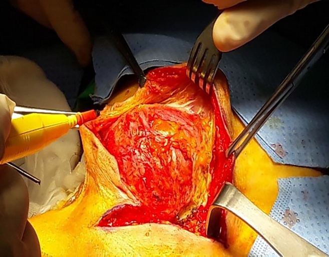
For the posterior approach (Fig. 3), patients were placed in a prone jack knife position and a straight paracoccygeal or inverted Y-shaped incision was usually made to just before the anal sphincter. Cysts were dissected with care taken to avoid perforating the rectum or cyst or leaving any cyst remnant. A digital rectal examination was performed intraoperatively to ensure rectal integrity. Large cysts at the coccyx or lower sacrum (S4–5) were removed with the help of orthopedic surgeons. All wounds were closed over negative suction drains.
For larger cysts, a combined approach was used starting with the laparoscopic anterior approach. After wound closure, patients were moved into the prone jack knife position for the posterior approach.
The choice of surgical approach was made according to tumor size and correlation with the pelvic floor. For infralevator tumors, the posterior approach was sufficient. For supralevator tumors, the anterior approach was necessary. For cysts with infralevator and supralevator extensions, if small (<5 cm), the posterior approach was sufficient. When cysts were 5–10 cm, the anterior approach was advised. Cysts >10 cm, may be multilocular with a dumbbell shape, so the combined approach was used.
Data analysis
Statistical analysis was accomplished with IBM SPSS Statistics ver. 23.0 (IBM Co., Armonk, NY, USA). Quantitative data were described as mean ± standard deviation. Qualitative data were presented as frequencies and proportions. Chi-square and 1-way analysis of variance tests were used. P-values < 0.05 were considered significant.
Our study included 24 patients who underwent surgical treatment for tailgut cyst that aimed for complete removal (Table 1). The median patient age was 51.5 years (range, 21–68 years). Of the patients 18 (75%) were females. Clinical presentations included pelvic pain in 4 patients, perineal pain in 3, palpable perianal swelling in 2, and a sacrococcygeal sinus with pus discharge posteriorly on the lower back in 1 patient. In the other 14 asymptomatic patients, cysts were incidentally discovered on radiology: 2 for urology problems; 4 for abdominal pain; 2 for chronic cholecystitis; 2 for gynecology problems; 4 via a medical checkup; and 2 via a colonoscopy, of which one was a rectal submucosal tumor.
For the 3 prerectal cysts, 1 was at the rectouterine pouch, 1 was at the rectovaginal septum, while the last one was in a male patient anterior to the rectum. The 3 prerectal tumors were below the level of the coccyx; all were at the supralevator level and managed via an anterior laparoscopic approach. An intraoperative vaginal injury occurred and was repaired in the 2 female patients. In the male patient, a rectal perforation occurred, for which repair and a loop ileostomy were performed. Postoperative courses were uneventful and patients were discharged home safely. The ileostomy was closed after 6 months in the male patient. Among the other 21 retrorectal cases, on MRI, tumors were below the level of the coccyx (coccyx down) in 13 and at the level of the coccyx in 5. Seven were managed using the anterior laparoscopic approach and most were supra- and infralevator. One female patient experienced a rectal perforation intraoperatively for whom ileostomy was performed. Her postoperative course was good, and she underwent ileostomy closure 2 months later. The posterior approach was used for the other 11 patients, who had tumors that mostly extended to the supralevator and infralevator levels. The other 3 patients (2 females, 1 male), on MRI, 2 had cysts extending above the level of the coccyx to the sacral promontory and 1 had a cyst opposite the coccyx; all were managed via the combined approach because of the large cyst size (10–14 cm) and dumbbell shape (Table 2).
Postoperative complications occurred only in 1 male patient (10%) in the anterior approach group and were Clavien-Dindo (CD) classification grade II (pelvic floor dyssynergia and lower limb weakness). The patient remained medicated without improvement. Complications occurred most frequently in the posterior approach group (P = 0.021) in comparison to other approaches. Wound seroma and infection (CD classification grade II) were the most common (7 patients, 63.6%), and all patients were managed with antibiotics and frequent dressing changes. One patient (9.1%) had postoperative fluid collection in the right ischiorectal fossa (CD classification grade III) that was managed by pigtail tube drainage. No postoperative complications occurred in the combined approach group except in that 1 male patient (33.3%) who had a posterior wound seroma and sexual dysfunction (CD classification grade II); he was under medical treatment.
Postoperative pathology findings were benign for all except 2 male patients (1 with carcinoid tumor, 1 with adenocarcinoma). The first patient was managed with radiotherapy, and the other was referred to the medical oncology staff, who recommended close follow-up as a sufficient safety margin was obtained in the lower sacrectomy and coccygectomy via the combined approach.
Median mass size was 5.2 cm (range, 2.3–14 cm). Median follow-up was 12 months (range, 1–66 months), with no recurrence was observed. No postoperative mortality occurred.
Patients treated with the combined approach had a significantly larger cyst size (P < 0.001), longer operation times (P < 0.001), and more tumors located above the coccyx (P = 0.002) than those treated using other approaches. No significant differences were noted for blood loss volume, time to first flatus, or tumor location relative to the pelvic floor muscles.
The incidence of retrorectal lesions is low in adults. A ratio of around 1/40,000 patients has been reported based on Mayo Clinic data [8]. Among retrorectal tumors, tailgut cysts arise from the remnants of the hind gut. Embryos normally form true tails, reaching maximum size at 35 days of gestation. The embryonic hindgut extends into the tail, forming the tailgut. By the week 8 of gestation, the tailgut normally regresses. Failure to obliterate this tailgut leads to the development of a tailgut cyst [9].
Tailgut cysts are rare and have a female predominance with a female to male ratio of 3 : 1 to 9 : 1. Patsouras and colleagues reported a female to male ratio of 7 : 1. Tailgut cysts commonly develop in the fifth decade of life; however, they can occur at any age [10-13]. In our study, the female to male ratio was 3 : 1 with a median patient age of 51.5 years.
Clinical presentation of tailgut cysts is usually nonspecific and misleading. Up to 50% of patients are asymptomatic, and cysts may be incidentally detected in radiological findings [13]. The other 50% of patients are symptomatic according to the largest reported case series of 53 patients by Hjermstad and Helwig [11]; symptoms usually occur from the local mass effect. Symptoms include rectal pain, constipation, obstructed defecation, tenesmus, painless rectal bleeding, dysuria, urinary frequency, lower abdominal pain, back pain, and lower limb neurological problems due to compression of the sacral plexus. Symptoms can also present as recurrent perianal suppuration, which is often misdiagnosed [11, 13]. In our series, 58.3% of patients were asymptomatic. Other patients’ symptoms included lower abdominal pain, perineal pain, perianal swelling and fistula, and sacrococcygeal sinus.
One of the valuable diagnostic tools for detecting unilocular, multilocular, and small peripheral cysts is pelvic MRI [14]. Cysts usually have low signal intensity on T1-weighted images and high signal intensity on T2-weighted images. However, high signal intensity can also be seen on T1-weighted images because of the presence of mucinous material, high protein content, or hemorrhage into the cyst [15-17]. Thus, with multiple capabilities and good tissue contrast, MRI is superior for surgical planning for presacral masses. MRI also gives insights into whether cysts are malignant. When malignant, cysts appear heterogeneous with solid and cystic content and irregular borders. However, recurrent benign cysts may also appear this way [14, 15]. In our study, all patients were diagnosed using MRI.
Controversy persists regarding preoperative biopsy. Hall et al. [18] demonstrated the possible use of ultrasound-guided needle biopsy as a diagnostic modality for tailgut cysts. Nevertheless, the use of this method is not advised as it carries the risk of seeding tumor cells [19]. Some patients experienced infection and tumor cell implantation when a biopsy sample was taken via transvaginal or transrectal (or, less commonly, para sacral) approaches. Some authors advised avoiding biopsy for masses that are purely cystic. For the cysts with mixed solid and cystic components, a percutaneous preoperative parasacral biopsy should be performed to determine treatment strategies with patients before surgery, optimize surgical plans, and inform patients about their prognosis and if postoperative adjuvant therapy is needed [12]. Others recommend performing biopsies only when complete excision is impossible [9, 12]. None of our patients underwent preoperative biopsy.
Tailgut cysts are usually lined with a variety of epithelia within the same cyst and may contain mucus; hence, malignant degeneration can occur within the cyst. Epithelial types are squamous, transitional, columnar, and cuboidal. Smooth muscle fibers are usually seen in association with cysts. Tailgut cyst may be associated with malignancy, mostly adenocarcinoma and carcinoid tumors. However, other reported tumors include neuroendocrine carcinoma, endometrioid carcinoma, adenosquamous carcinoma, squamous cell carcinoma, and sarcoma [11, 19, 20]. Our study had 2 patients with malignancy (both male: adenocarcinoma and carcinoid tumor). No females had malignant cysts. However, Mathis et al. [12] reported 4 female patients with malignancy (3 adenocarcinomas and 1 carcinoid tumor).
Studies reported a different incidence for malignant transformation of tailgut cysts. The rate of malignant transformation in the largest series in the literature in 1988 was 2% [10]. Patsouras et al. [13] reported a 6% rate of malignant degeneration, other studies reported 13 to 40% [12, 14], and in our study, the rate was 8%.
Differential diagnosis should always be considered for retrorectal cysts. These are dermoid, epidermoid cyst, rectal duplication cyst, neuroenteric cyst, teratoma, anterior sacral meningocele, leiomyosarcoma, cystic lymphangioma, pyogenic abscess, neurogenic cyst, sacral chordoma, and tailgut cyst [19]. For prerectal cysts, the differential diagnosis includes prostatic utricle, simple cyst of the prostate, rectal duplication, simple cyst of the seminal vesicle, and bladder diverticulum [21].
The choice of surgical approach depends on several factors, including the degree of proximal extension of the cyst, cyst size, presence of infection, adherence to neighboring structures (bladder, ureters, rectum), and absence or presence of evidence of malignancy necessitating en bloc resection [12, 13]. The posterior approach is most frequently described [22, 23].
For prerectal cysts, caution and careful dissection should be used to avoid injury to adjacent structures including the vagina, uterus, bladder, and rectum itself, especially when the cyst adheres to them. Injury occurred in our 3 prerectal cases with, 2 vaginal and 1 rectal. In our study, the risk of intra operative complications was high for prerectal compared to the retrorectal cysts.
Managing tailgut cysts via a trans rectal approach has been reported by some studies, most of which are case reports. McCarroll and Moore [24] reported managing a patient via a transanal minimally invasive surgery technique with no postoperative complications. Kildušis and Samalavičius [25] reported a patient managed by an open transrectal approach, also without complications. This study, noted that the transrectal approach can be for small noninfected low-lying cysts.
Debate persists about whether the coccyx should be removed with the cyst. Several studies advocate coccygectomy since it improves surgical exposure and decreases risk of recurrence assuming that the coccyx may harbor a nidus of totipotential cellular remnants that may cause recurrence [4, 26]. However, the concern that the coccyx will increase risk of recurrence has not been demonstrated in other studies, especially for cysts not adherent to the coccyx and that can be removed entirely without coccygectomy; hence, coccyx removal is not required [27]. Removal of coccyx was based on the cyst itself and not the coccyx harboring aberrant remnants of the postanal gut leading to cyst formation. Recent trials report preserving the coccyx unless en bloc resection is required for malignancy or cysts densely adhere to the coccyx. Based on our findings, we removed the coccyx for 4 patients: 3 due to the cyst adhering to the coccyx and 1 for a large cyst requiring a combined coccygectomy and lower sacrectomy; the latter cyst was malignant.
We noted the importance of dissection in the proper surgical plane to avoid injury to the hypogastric nerves and pelvic plexuses during the anterior approach. Mathis et al. [12] reported complications of pelvic floor dyssynergia in 2 patients whose conditions improved with biofeedback therapy and 1 patient with sexual dysfunction after reoperation for recurrence. In our study, 1 patient had sexual dysfunction after the combined approach and 1 patient had pelvic floor dyssynergia and lower limb weakness after treatment with the anterior approach; both remain under medical treatment and follow-up. Other complications including postoperative wound seroma and infection were frequently observed after the posterior approach in our study; all were managed conservatively.
Avoiding injury to the rectal wall during dissection of the tailgut cyst is important using the posterior approach. Injury avoidance is facilitated by applying soft paraffin gauze roll packing in the rectum as a guide for rectum identification together with the preoperative bowel preparation.
Limited reports exist on recurrence rates after tailgut cyst resection. Some reported a range of 0%–16% and usually due to incomplete excision [13]. Another series found no recurrence among 12 patients after a median follow-up 54 months [3]. Mathias et al. [12] reported 1 case of recurrence after 5 years of followup. We had no cases of recurrence after a median follow-up of 12 months.
For surgical timing, some studies suggest routine follow-up for a symptomatic patient with low malignancy risk. may be appropriate. However, a definitive diagnosis cannot be obtained except after surgical excision and histopathological examination [28]. Accordingly, surgery is mandatory to exclude malignancy.
Despite being retrospective, our study included a large number of patients. The anterior approach and anterior part of the combined approach were performed laparoscopically in all cases, and no recurrence occurred after a median 12-month follow-up. We provide a management strategy according to tumor size and its correlation to pelvic floor muscles.
In conclusion, despite being uncommon, tailgut cysts can cause compression manifestations such as perineal or pelvic pain. Surgical excision is mandatory regardless of symptom status because of the risk of malignancy. Complete surgical excision is a key treatment using an appropriate approach according to tumor size, location, and correlation with adjacent pelvic floor muscles.

CONFLICT OF INTEREST

The authors report no potential conflicts of interest relevant to this article.

Fig. 1.
(A) Malignant cyst extending above the coccyx showing soft tissue component inside. (B) The arrow pointing at the cyst opposite the level of the coccyx. (C) The arrow pointing at the cyst below the level of the coccyx. (D) Dumbbell shaped cyst.
ac-2018-12-18f1.jpg
Fig. 2.
Laparoscopic anterior approach view.
ac-2018-12-18f2.jpg
Fig. 3.
Posterior approach view.
ac-2018-12-18f3.jpg
Table 1.
Patient demographics
Characteristic Value
Age (yr) 51.5 (21.0–68.0)
Sex
 Male 6 (25.0)
 Female 18 (75.0)
Symptoms
 Asymptomatic 14 (58.3)
 Lower abdominal pain 4 (16.7)
Perineal pain 3 (12.5)
 Palpable perianal swelling 1 (4.2)
 Perianal abscess 1 (4.2)
Sacrococcygeal sinus 1 (4.2)
Mass size (cm) 5.2 (2.3–14.0)
Tumor location
 Prerectal 3 (12.5)
 Retrorectal 21 (87.5)
Tumor level related to coccyx
 Below 16 (66.7)
 Opposite 6 (25.0)
 Above 2 (8.3)
Tumor level related to pelvic floor
 Supralevator 5 (20.8)
 Supra- + infralevator 18 (75.0)
 Infralevator 1 (4.2)
Approach
 Anterior, laparoscopic 10 (41.7)
 Posterior 11 (45.8)
 Combined 3 (12.5)
Intraoperative complication
 Vaginal injury 2 (8.3)
 Rectal serosal injury 2 (8.3)
 Rectal injury requiring diversion 2 (8.3)
Postoperative complication
 Wound seroma and infection 7 (29.2)
 Fluid collection in ischiorectal fossa 1 (4.2)
 Lower limb weakness, pelvic floor dyssynergia 1 (4.2)
 Sexual dysfunction 1 (4.2)
Clavien-Dindo classification grade
 I 0 (0)
 II 9 (37.5)
 III 1 (4.2)
 IV 0 (0)
 V 0 (0)
Pathology
 Benign 22 (91.7)
 Malignanta 2 (8.3)
Follow-up (mo) 12 (1–66)
Recurrence 0 (0)
Mortality 0 (0)

Values are presented as median (range) or number (%).

aOne adenocarcinoma with close follow-up, 1 carcinoid with radiotherapy.

Table 2.
Comparison between the 3 surgical approaches
Variable Anterior approach (n = 10) Posterior approach (n = 11) Combined approach (n = 3) P-value
Coccyx 0.002
 Above 0 (0) 0 (0) 2 (66.7)
 Opposite 3 (30) 2 (18.2) 1 (33.3)
 Belowa 7 (70) 9 (81.8) 0 (0)
Pelvic floor 0.296
 Supralevatora 4 (40) 1 (9.1) 0 (0)
 Supra- + infralevator 6 (60) 9 (81.8) 3 (100)
 Infralevator 0 (0) 1 (9.1) 0 (0)
 Size (cm) 5.2 ± 1.6 4.3 ± 1.3 11.4 ± 2.3 0.001
 EBL (mL) 35 (0–750) 20 (0–500) 200 (100–700) 0.149
 Operative time (min) 157 (100–360) 90 (45–200) 320 (315–400) 0.001
 Time to first flatus (day) 1.8 ± 1.1 1.1 ± 0.3 2.0 ± 1.00 0.075
 Clavien-Dindo classification grade 0.021
  I 0 (0) 0 (0) 1 (33.3)
  II 1 (10) 7 (63.6) 0 (0)
  III 0 (0) 1 (9.1) 0 (0)
  IV 0 (0) 0 (0) 0 (0)
  V 0 (0) 0 (0) 0 (0)

Values are presented as number (%), mean ± standard deviation, or median (range).

EBL, estimated blood loss.

aLocation of 3 cases of prerectal tumors.

  • 1. Vega Menéndez D, Quintáns Rodríguez A, Hernández Granados P, Nevado Santos M, García Sabrido JL, Rueda Orgaz JA, et al. Tailgut cysts. Cir Esp 2008;83:53–60.ArticlePubMed
  • 2. Dahan H, Arrivé L, Wendum D, Docou le Pointe H, Djouhri H, Tubiana JM. Retrorectal developmental cysts in adults: clinical and radiologic-histopathologic review, differential diagnosis, and treatment. Radiographics 2001;21:575–84.ArticlePubMed
  • 3. Lev-Chelouche D, Gutman M, Goldman G, Even-Sapir E, Meller I, Issakov J, et al. Presacral tumors: a practical classification and treatment of a unique and heterogeneous group of diseases. Surgery 2003;133:473–8.ArticlePubMed
  • 4. Wolpert A, Beer-Gabel M, Lifschitz O, Zbar AP. The management of presacral masses in the adult. Tech Coloproctol 2002;6:43–9.ArticlePubMedPDF
  • 5. Gips M, Melki Y, Wolloch Y. Cysts of the tailgut. Two cases. Eur J Surg 1994;160:459–60.PubMed
  • 6. Sidoni A, Bucciarelli E. Ciliated cyst of the perineal skin. Am J Dermatopathol 1997;19:93–6.ArticlePubMed
  • 7. Sung MT, Ko SF, Niu CK, Hsieh CS, Huang HY. Perirenal tailgut cyst (cystic hamartoma). J Pediatr Surg 2003;38:1404–6.ArticlePubMed
  • 8. Jao SW, Beart RW Jr, Spencer RJ, Reiman HM, Ilstrup DM. Retrorectal tumors. Mayo Clinic experience, 1960-1979. Dis Colon Rectum 1985;28:644–52.ArticlePubMed
  • 9. Prasad AR, Amin MB, Randolph TL, Lee CS, Ma CK. Retrorectal cystic hamartoma: report of 5 cases with malignancy arising in 2. Arch Pathol Lab Med 2000;124:725–9.PubMed
  • 10. Killingsworth C, Gadacz TR. Tailgut cyst (retrorectal cystic hamartoma): report of a case and review of the literature. Am Surg 2005;71:666–73.ArticlePubMed
  • 11. Hjermstad BM, Helwig EB. Tailgut cysts. Report of 53 cases. Am J Clin Pathol 1988;89:139–47.ArticlePubMedPDF
  • 12. Mathis KL, Dozois EJ, Grewal MS, Metzger P, Larson DW, Devine RM. Malignant risk and surgical outcomes of presacral tailgut cysts. Br J Surg 2010;97:575–9.ArticlePubMed
  • 13. Patsouras D, Pawa N, Osmani H, Phillips RK. Management of tailgut cysts in a tertiary referral centre: a 10-year experience. Colorectal Dis 2015;17:724–9.ArticlePubMed
  • 14. Yang DM, Park CH, Jin W, Chang SK, Kim JE, Choi SJ, et al. Tailgut cyst: MRI evaluation. AJR Am J Roentgenol 2005;184:1519–23.ArticlePubMed
  • 15. Kim MJ, Kim WH, Kim NK, Yun MJ, Park YN, Lee JT, et al. Tailgut cyst: multilocular cystic appearance on MRI. J Comput Assist Tomogr 1997;21:731–2.ArticlePubMed
  • 16. Lim KE, Hsu WC, Wang CR. Tailgut cyst with malignancy: MR imaging findings. AJR Am J Roentgenol 1998;170:1488–90.ArticlePubMed
  • 17. Moulopoulos LA, Karvouni E, Kehagias D, Dimopoulos MA, Gouliamos A, Vlahos L. MR imaging of complex tail-gut cysts. Clin Radiol 1999;54:118–22.ArticlePubMed
  • 18. Hall DA, Pu RT, Pang Y. Diagnosis of foregut and tailgut cysts by endosonographically guided fine-needle aspiration. Diagn Cytopathol 2007;35:43–6.ArticlePubMed
  • 19. Gönül II, Bağlan T, Pala I, Menteş B. Tailgut cysts: diagnostic challenge for both pathologists and clinicians. Int J Colorectal Dis 2007;22:1283–5.ArticlePubMedPDF
  • 20. Song DE, Park JK, Hur B, Ro JY. Carcinoid tumor arising in a tailgut cyst of the anorectal junction with distant metastasis: a case report and review of the literature. Arch Pathol Lab Med 2004;128:578–80.ArticlePubMedPDF
  • 21. Jang SH, Jang KS, Song YS, Min KW, Han HX, Lee KG, et al. Unusual prerectal location of a tailgut cyst: a case report. World J Gastroenterol 2006;12:5081–3.ArticlePubMedPMC
  • 22. Hannon J, Subramony C, Scott-Conner CE. Benign retrorectal tumors in adults: the choice of operative approach. Am Surg 1994;60:267–72.PubMed
  • 23. Singer MA, Cintron JR, Martz JE, Schoetz DJ, Abcarian H. Retrorectal cyst: a rare tumor frequently misdiagnosed. J Am Coll Surg 2003;196:880–6.ArticlePubMed
  • 24. McCarroll RH, Moore LJ. Transanal minimally invasive surgery for resection of retrorectal cyst. J Surg Case Rep 2018;2018:rjy021. ArticlePubMedPMCPDF
  • 25. Kildušis E, Samalavičius NE. Surgical management of a retro-rectal cystic hamartoma (tailgut cyst) using a trans-rectal approach: a case report and review of the literature. J Med Case Rep 2014;8:11. ArticlePubMedPMCPDF
  • 26. Izant RJ Jr, Filston HC. Sacrococcygeal teratomas. Analysis of forty-three cases. Am J Surg 1975;130:617–21.ArticlePubMed
  • 27. Böhm B, Milsom JW, Fazio VW, Lavery IC, Church JM, Oakley JR. Our approach to the management of congenital presacral tumors in adults. Int J Colorectal Dis 1993;8:134–8.ArticlePubMedPDF
  • 28. Au E, Anderson O, Morgan B, Alarcon L, George ML. Tailgut cysts: report of two cases. Int J Colorectal Dis 2009;24:345–50.ArticlePubMedPDF

Figure & Data

References

    Citations

    Citations to this article as recorded by  
    • Risk factors for rectal perforation during presacral cyst removal: a comparison between transabdominal, perineal, and combined surgical approaches
      P. Tsarkov, S. Barkhatov, D. Shlyk, L. Safyanov, V. Balaban, M. He
      Techniques in Coloproctology.2025;[Epub]     CrossRef
    • Tailgut Cyst—Gynecologist’s Pitfall: Literature Review and Case Report
      Andrei Mihai Malutan, Viorela-Elena Suciu, Florin Laurentiu Ignat, Doru Diculescu, Razvan Ciortea, Emil-Claudiu Boțan, Carmen Elena Bucuri, Maria Patricia Roman, Ionel Nati, Cristina Ormindean, Dan Mihu
      Diagnostics.2025; 15(1): 108.     CrossRef
    • Laparoscopic Excision of a Tailgut Cyst With Refractory Pain: A Case Report
      Masato Kitazawa, Seishu Karasawa, Satoshi Nakamura, Yuta Yamamoto, Yuji Soejima
      Cureus.2025;[Epub]     CrossRef
    • Diagnostic challenges of tailgut cysts: a case report on an occult perianal mass
      Faraz Khalid, Haris Naveed, Masab Ali, Muhammad Saad Ansari, Muhammad Sajjad Shafiq, Muhammad Husnain Ahmad
      Annals of Medicine & Surgery.2025; 87(1): 421.     CrossRef
    • Transanal-transrektale endoskopische Resektion einer Schwanzdarmzyste
      Hussein Abdallah, Stefan Heinrich, Matthias Birth
      coloproctology.2025; 47(3): 220.     CrossRef
    • Partial Resection of a Tailgut Cyst Attached to the Rectum via a Transcoccygeal Approach: A Case Report With a Favorable Outcome
      Eitaro Okumura, Motoo Kubota, Ouji Momosaki, Ryo Hashimoto, Kotaro Kohara
      Cureus.2025;[Epub]     CrossRef
    • Recurrent presacral tailgut cyst with mucinous adenocarcinoma and metastasis
      Iyad Al Jada, Majd Oweidat, Mohammad Khaleel, Omar Harb, Ursula Abu Nahla, Rahaf Bleibel, Mai Arafeh, Ammar W.M. Hassouneh
      Radiology Case Reports.2025; 20(8): 4022.     CrossRef
    • Trans‐abdominal single‐incision robotic surgery with the da Vinci SP® surgical system for 8 cases of retrorectal tumour
      Ho Seung Kim, Bo‐Young Oh, Soon Sup Chung, Ryung‐Ah Lee, Gyoung Tae Noh
      The International Journal of Medical Robotics and Computer Assisted Surgery.2024;[Epub]     CrossRef
    • Three Cases of Anal Cystic Lesions in Our Hospital
      Kaori Tanaka, Toshiharu Mori, Hideki Yamada, Hideki Mori
      Nihon Daicho Komonbyo Gakkai Zasshi.2024; 77(4): 199.     CrossRef
    • Diagnosis and treatment of retrorectal cystic hamartoma: clinical case
      S. A. Zvezda, D. G. Dimitriadi, P. I. Tamrazov, A. V. Simonov, E. M. Frank, N. M. Fedorov, L. N. Komarova
      Surgery and Oncology.2024; 14(1): 72.     CrossRef
    • Presacral Tailgut Cyst
      Shriya Haval, Divyansh Dwivedi, Prabhat Nichkaode
      Annals of African Medicine.2024; 23(2): 237.     CrossRef
    • A neuroendocrine tumor arising in a tailgut cyst: Case report and literature review
      Wei Guo, Ming Deng, Qiongrong Chen
      International Journal of Surgery Case Reports.2024; 120: 109912.     CrossRef
    • Tailgut Cyst in a Child: A Case Report and Review of Literature
      Ramendra Shukla, Jay Divyesh Patel, Sudhir B. Chandna, Urvish Parikh
      African Journal of Paediatric Surgery.2024; 21(3): 184.     CrossRef
    • Two Cases of Symptomatic Tailgut Cysts
      Jan Wojciechowski, Tomasz Skołozdrzy, Piotr Wojtasik, Maciej Romanowski
      Journal of Clinical Medicine.2024; 13(17): 5136.     CrossRef
    • The management of retrorectal tumors – a single-center analysis of 21 cases and overview of the literature
      K. Fechner, B. Bittorf, M. Langheinrich, K. Weber, M. Brunner, R. Grützmann, K. E. Matzel
      Langenbeck's Archives of Surgery.2024;[Epub]     CrossRef
    • Kraske-Zugang für Tumoren im kaudalen retrorektalen Raum
      Rosita Sortino, Sebastian Christen, Daniel Steinemann, Ida Füglistaler
      coloproctology.2024; 46(6): 413.     CrossRef
    • Laparoscopic approach in the surgical treatment of large retrorectal tumors: a short-term experience at a single tertiary center case series in Korea
      Jun Seong Chung, Han Deok Kwak, Jae Kyun Ju
      Journal of Minimally Invasive Surgery.2024; 27(4): 221.     CrossRef
    • Open surgical treatment for giant presacral tailgut cyst – a case report
      Q Liu, P Guo, C Li, G Yu, J Jiao
      South African Journal of Surgery.2024; 62(4): 358.     CrossRef
    • Cystic lesions of the retrorectal space
      Ian S Brown, Anna Sokolova, Christophe Rosty, Rondell P Graham
      Histopathology.2023; 82(2): 232.     CrossRef
    • Tailgut cyst mimicking second anal opening in an infant
      Jay Lodhia, Mujaheed Suleman, Doreen Msemakweli, Joshua Tadayo, Patrick Amsi, David Msuya
      Journal of Pediatric Surgery Case Reports.2023; 89: 102550.     CrossRef
    • Surgery of presacral cysts: case series analysis and literature review
      K. V. Stegnii, Zh. A. Rakhmonov, R. A. Goncharuk, M. A. Krekoten, E. R. Dvoinikova, E. V. Morova, M. О. Dmitriev
      Pacific Medical Journal.2023; (2): 15.     CrossRef
    • Surgical treatment of retrorectal tumors: a plea for a laparoscopic approach
      Clara Galán, M. Pilar Hernández, M. Carmen Martínez, Anna Sánchez, Jesús Bollo, Eduardo Mª Targarona
      Surgical Endoscopy.2023; 37(12): 9080.     CrossRef
    • Robotic approach to remove four tailgut cyst cases in Brazil: a case series
      Bruno Mirandola Bulisani, Luiz Guilherme Lisboa Gomes, Milena Arruda de Oliveira Leite, Ricardo Moreno, Murilo Rocha Rodrigues, Felipe Martin Bianco Rossi, Renato Barretto Ferreira da Silva, Luiz Carlos Benjamin do Carmo, Jaques Waisberg
      einstein (São Paulo).2023;[Epub]     CrossRef
    • Tumor neuroendocrino asociado con quiste intestinal: presentación de un caso
      Lismary Ruiz Cabezas, Alicia Moreno Ontalba, Mario Díaz Delgado, Esther María Cidoncha Pérez, Alejandro Rubio Fernández, María Victoria González Ibáñez
      Revista Española de Patología.2022; 55(4): 278.     CrossRef
    • Risk of malignancy and outcomes of surgically resected presacral tailgut cysts: A current review of the Mayo Clinic experience
      Sacha P. Broccard, Dorin T. Colibaseanu, Kevin T. Behm, Nitin Mishra, Peter Davis, Kristi L. Maimone, Kellie L. Mathis, Luca Stocchi, Eric J. Dozois, Amit Merchea
      Colorectal Disease.2022; 24(4): 422.     CrossRef
    • Squamous cell carcinoma in a retrorectal cystic hamartoma
      Quintáns Ana Teijo, Valadés José Ignacio Martín , del Río Cristina Garrán , Rojo Irene López , Casado Oscar Alonso
      Journal of Radiology and Oncology.2022; 6(1): 001.     CrossRef
    • Retrorectal Tailgut Cyst: A Case Report
      Neel Shah, Peter Edelstein
      Cureus.2022;[Epub]     CrossRef
    • A Case of Tailgut Cyst Treated with Laparoscopic Transabdominal and Transsacral Surgery
      Naoki Kawahara, Yoshiyuki Suzuki, Yu Sakai, Shinji Maeshima, Takehiro Shimada
      Nippon Daicho Komonbyo Gakkai Zasshi.2022; 75(4): 170.     CrossRef
    • Promoting Laparoscopic Anterior Approach for a Very Low Presacral Primary Neuroendocrine Tumor Arising in a Tailgut Cyst
      Maria Michela Di Nuzzo, Carlo De Werra, Mirella Pace, Raduan Ahmed Franca, Maria D’Armiento, Umberto Bracale, Ruggero Lionetti, Michele D’Ambra, Armando Calogero
      Healthcare.2022; 10(5): 805.     CrossRef
    • A Case of Dermoid Cyst in the Ischiorectal Fossa Resected by the Transperineal Approach
      Susumu Daibo, Hirokazu Suwa, Seiya Sato, Koki Goto, Yusuke Suwa, Jun Watanabe
      Nippon Daicho Komonbyo Gakkai Zasshi.2022; 75(5): 237.     CrossRef
    • Perianal Tailgut Cyst: An Unusual Presentation
      Rabia Arshad, Noor Khalid, Mubashir Rafique, Ruqia Mushtaq, Fakhar Munir Sial
      Cureus.2022;[Epub]     CrossRef
    • Squamous cell carcinoma arising in a tailgut cyst: role of radiotherapy
      Diego Aldave, Ana Teijo, Claudia Abril, Laura Cerezo
      BMJ Case Reports.2022; 15(9): e247985.     CrossRef
    • Hamartoma quístico retrorrectal (tailgut cyst): reporte de un caso y revisión de la literatura
      Juan Dario Puerta Diaz, Rodrigo Castaño Llano, Alfredo Enrique Martelo Román, Juan Esteban Puerta Botero
      Revista colombiana de Gastroenterología.2022; 37(3): 316.     CrossRef
    • Diagnostic, Therapeutic and Evolutionary Problems of Retro-Rectal Tumors (About 3 Cases)
      Hajri Amal, Ahmed Elmi Abdirahim, Rachid Boufettal, Erguibi Driss, Saad Rifki El Jai, Farid Chehab
      European Journal of Medical and Health Sciences.2022; 4(5): 13.     CrossRef
    • Epidemiology, diagnostic approach and therapeutic management of tailgut cysts: A systematic review
      Aikaterini Mastoraki, Ilias Giannakodimos, Karmia Panagiotou, Maximos Frountzas, Dimosthenis Chrysikos, Stylianos Kykalos, Georgios E. Theodoropoulos, Dimitrios Schizas
      International Journal of Clinical Practice.2021;[Epub]     CrossRef
    • Re: Malignant transformation of tailgut cysts is significantly higher than previously reported: Systematic review of cases in the literature
      Tamara Glyn, Peter Sagar, Frank Frizelle
      Colorectal Disease.2021; 23(11): 3027.     CrossRef
    • Multicenter Analysis of Presacral Neuroendocrine Neoplasms—Clinicopathological Characterization and Treatment Outcomes of a Rare Disease
      Sami Matrood, Leonidas Apostolidis, Jörg Schrader, Sebastian Krug, Harald Lahner, Annette Ramaswamy, Damiano Librizzi, Zoltan Kender, Anke Kröcher, Simon Kreutzfeldt, Thomas Matthias Gress, Anja Rinke
      Frontiers in Endocrinology.2021;[Epub]     CrossRef
    • Surgical Management of Retrorectal Tumors
      Mathilde Aubert, Diane Mege, Yann Parc, Eric Rullier, Eddy Cotte, Guillaume Meurette, Philippe Zerbib, Bertrand Trilling, Bernard Lelong, Charles Sabbagh, Zaher Lakkis, Mehdi Ouaissi, Gil Lebreton, Philippe Rouanet, Gilles Manceau, Jean-Jacques Tuech, Gui
      Annals of Surgery.2021; 274(5): 766.     CrossRef
    • Isolated lumbar intradural tailgut cyst: A case report and review of the literature
      Antonio Colamaria, Matteo Sacco, Giovanni Parbonetti, Maria Blagia, Francesco Carbone, Matteo de Notaris
      Heliyon.2021; 7(10): e08223.     CrossRef
    • Prerectal tailgut cyst presenting with constipation and pseudo-rectal prolapse. A rare case presentation
      Allan Ngulube, Blessing Zambuko, Crispin O.M. Ntoto, Simbarashe Gift Mungazi, Derek Matsika
      Human Pathology Reports.2021; 26: 300557.     CrossRef
    • Anal Canal Duplication in an Adult Female—Case Report and Pathology Guiding
      Tudor Mateescu, Cristi Tarta, Paul Stanciu, Alis Dema, Fulger Lazar
      Medicina.2021; 57(11): 1205.     CrossRef
    • “The Innate Tail”: An Unusual Cause of Rectal Bleeding in an Adult Male
      Sujit Padmanabhan Nair, Sanjay Chandnani, Prasanta Debnath, Pravin M. Rathi, Parmeshwar Junare, Vinay Zanwar, Sangeeta Kini
      GE - Portuguese Journal of Gastroenterology.2021; 28(5): 349.     CrossRef
    • Robotic approach to large tailgut cyst with malignant transformation: A case report
      Alessandra Marano, Maria Carmela Giuffrida, Chiara Peluso, Valentina Testa, Paolo Bosio, Felice Borghi
      International Journal of Surgery Case Reports.2020; 77: S57.     CrossRef
    • Laparoscopic resection of a neuroendocrine tumor that almost fully replaced tailgut cysts: a case report
      Keita Kodera, Seiichiro Eto, Nei Fukasawa, Wataru Kai, Tomo Matsumoto, Tsuyoshi Hirabayashi, Hidejiro Kawahara, Nobuo Omura
      Surgical Case Reports.2020;[Epub]     CrossRef
    • Retrorectal tumors: Case report and review of literature
      Giorgio La Greca, Giovanni Trombatore, Guido Basile, Pietro Conti
      International Journal of Surgery Case Reports.2020; 77: 726.     CrossRef
    • A Tailgut Cyst in the Ischiorectal Fossa—A Case Report—
      Ryo INADA, Eri KURODA, Ayako WATANABE, Toshiaki TOSHIMA, Kazuhide OZAKI, Yuichi SHIBUYA, Manabu MATSUMOTO, Jun IWATA
      Nihon Rinsho Geka Gakkai Zasshi (Journal of Japan Surgical Association).2020; 81(9): 1866.     CrossRef
    • Tailgut Cyst, Report of 24 Cases Single Center Experience
      Han Deok Kwak, Chang Hyun Kim
      Annals of Coloproctology.2019; 35(5): 227.     CrossRef

    • Cite this Article
      Cite this Article
      export Copy Download
      Close
      Download Citation
      Download a citation file in RIS format that can be imported by all major citation management software, including EndNote, ProCite, RefWorks, and Reference Manager.

      Format:
      • RIS — For EndNote, ProCite, RefWorks, and most other reference management software
      • BibTeX — For JabRef, BibDesk, and other BibTeX-specific software
      Include:
      • Citation for the content below
      Single-center Experience of 24 Cases of Tailgut Cyst
      Ann Coloproctol. 2019;35(5):268-274.   Published online October 31, 2019
      Close
    • XML DownloadXML Download
    Figure
    • 0
    • 1
    • 2
    Single-center Experience of 24 Cases of Tailgut Cyst
    Image Image Image
    Fig. 1. (A) Malignant cyst extending above the coccyx showing soft tissue component inside. (B) The arrow pointing at the cyst opposite the level of the coccyx. (C) The arrow pointing at the cyst below the level of the coccyx. (D) Dumbbell shaped cyst.
    Fig. 2. Laparoscopic anterior approach view.
    Fig. 3. Posterior approach view.
    Single-center Experience of 24 Cases of Tailgut Cyst
    Characteristic Value
    Age (yr) 51.5 (21.0–68.0)
    Sex
     Male 6 (25.0)
     Female 18 (75.0)
    Symptoms
     Asymptomatic 14 (58.3)
     Lower abdominal pain 4 (16.7)
    Perineal pain 3 (12.5)
     Palpable perianal swelling 1 (4.2)
     Perianal abscess 1 (4.2)
    Sacrococcygeal sinus 1 (4.2)
    Mass size (cm) 5.2 (2.3–14.0)
    Tumor location
     Prerectal 3 (12.5)
     Retrorectal 21 (87.5)
    Tumor level related to coccyx
     Below 16 (66.7)
     Opposite 6 (25.0)
     Above 2 (8.3)
    Tumor level related to pelvic floor
     Supralevator 5 (20.8)
     Supra- + infralevator 18 (75.0)
     Infralevator 1 (4.2)
    Approach
     Anterior, laparoscopic 10 (41.7)
     Posterior 11 (45.8)
     Combined 3 (12.5)
    Intraoperative complication
     Vaginal injury 2 (8.3)
     Rectal serosal injury 2 (8.3)
     Rectal injury requiring diversion 2 (8.3)
    Postoperative complication
     Wound seroma and infection 7 (29.2)
     Fluid collection in ischiorectal fossa 1 (4.2)
     Lower limb weakness, pelvic floor dyssynergia 1 (4.2)
     Sexual dysfunction 1 (4.2)
    Clavien-Dindo classification grade
     I 0 (0)
     II 9 (37.5)
     III 1 (4.2)
     IV 0 (0)
     V 0 (0)
    Pathology
     Benign 22 (91.7)
     Malignanta 2 (8.3)
    Follow-up (mo) 12 (1–66)
    Recurrence 0 (0)
    Mortality 0 (0)
    Variable Anterior approach (n = 10) Posterior approach (n = 11) Combined approach (n = 3) P-value
    Coccyx 0.002
     Above 0 (0) 0 (0) 2 (66.7)
     Opposite 3 (30) 2 (18.2) 1 (33.3)
     Belowa 7 (70) 9 (81.8) 0 (0)
    Pelvic floor 0.296
     Supralevatora 4 (40) 1 (9.1) 0 (0)
     Supra- + infralevator 6 (60) 9 (81.8) 3 (100)
     Infralevator 0 (0) 1 (9.1) 0 (0)
     Size (cm) 5.2 ± 1.6 4.3 ± 1.3 11.4 ± 2.3 0.001
     EBL (mL) 35 (0–750) 20 (0–500) 200 (100–700) 0.149
     Operative time (min) 157 (100–360) 90 (45–200) 320 (315–400) 0.001
     Time to first flatus (day) 1.8 ± 1.1 1.1 ± 0.3 2.0 ± 1.00 0.075
     Clavien-Dindo classification grade 0.021
      I 0 (0) 0 (0) 1 (33.3)
      II 1 (10) 7 (63.6) 0 (0)
      III 0 (0) 1 (9.1) 0 (0)
      IV 0 (0) 0 (0) 0 (0)
      V 0 (0) 0 (0) 0 (0)
    Table 1. Patient demographics

    Values are presented as median (range) or number (%).

    One adenocarcinoma with close follow-up, 1 carcinoid with radiotherapy.

    Table 2. Comparison between the 3 surgical approaches

    Values are presented as number (%), mean ± standard deviation, or median (range).

    EBL, estimated blood loss.

    Location of 3 cases of prerectal tumors.


    Ann Coloproctol : Annals of Coloproctology Twitter Facebook
    TOP