


INTRODUCTION
A renal leiomyosarcoma is an uncommon neoplasm of the kidney, accounting for approximately 1% of all malignant renal tumors, and has a poor prognosis [1, 2]. It usually remains asymptomatic until the tumor grows to a relatively large size. Its symptoms, such as nonspecific abdominal discomfort, dull pain, and oliguria, are common in other conditions; therefore, a renal leiomyosarcoma is difficult to diagnose preoperatively. In this case, the condition had been preoperatively misdiagnosed four years earlier as a renal cell carcinoma before it was diagnosed as a primary renal leiomyosarcoma. Furthermore, it took several months to diagnose a recurrent renal leiomyosarcoma in the same patient because of the ambiguous, nonspecific symptoms. She visited our clinic complaining of diffuse abdominal pain, and colonoscopy showed a submucosal tumor or an extrinsic mass involving the descending colon. There are no reported cases of a renal leiomyosarcoma invading the retroperitoneal colon and mimicking a submucosal lesion. We report this case and present a literature review.
CASE REPORT
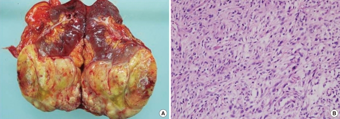
A 57-year-old woman visited our gastroenterology department complaining of diffuse lower abdominal discomfort. She had undergone a left nephrectomy four years earlier. The preoperative diagnosis was a renal cell carcinoma, but she was subsequently diagnosed with a primary renal leiomyosarcoma on pathologic examination (Fig. 1). Regular follow-ups with a urologist and abdominal computed tomography (CT) showed no recurrence for three years. After another year, she returned to the hospital complaining of diffuse abdominal discomfort. At that time, she was taking medication for hypertension and diabetes mellitus.
The findings of a general physical examination were unremarkable, except for diffuse abdominal sensitivity. The laboratory test results were as follows: leukocyte count, 6,770/mm3; hemoglobin level, 12.0 g/dL; and platelet count, 276,000/mm3. The levels of aspartate aminotransferase (12 IU/L), alanine transaminase (10 IU/L), alkaline phosphatase (82 IU/L), and total bilirubin (1.2 mg/dL) were within the reference range.
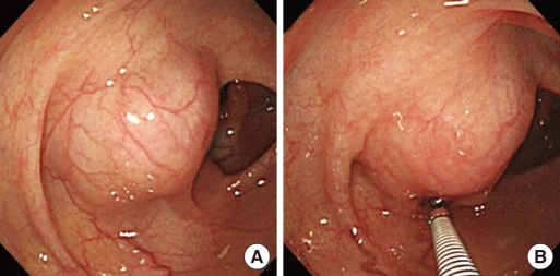
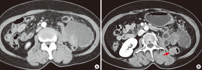
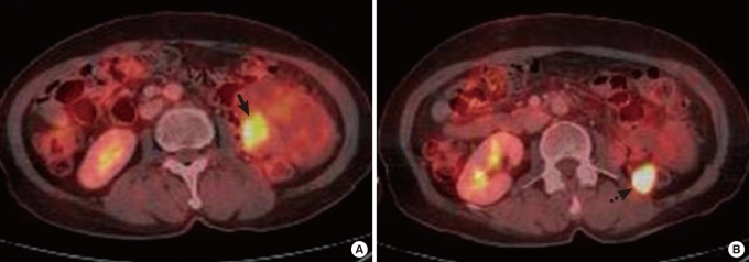
Colonoscopy revealed a hemispheric lesion similar to a submucosal tumor in the descending colon, approximately 30 cm above the anal verge (Fig. 2). The mass was found to be hard and fixed. An abdominal CT scan showed two mass lesions invading the descending colon (Fig. 3). The larger lesion was located in the peritoneum, adjacent to the anterior portion of the descending colon, and the smaller lesion was located in the retroperitoneum, adjacent to the posterior portion of the descending colon. Positron emission tomography-computed tomography (PET-CT) was performed. PET-CT showed a large omental mass on the left side of the abdomen, with a small solid area of increased glucose metabolism and hypermetabolism (maximum standardized uptake value of 13.7), in the posteromedial portion of the mid-descending colon (Fig. 4).
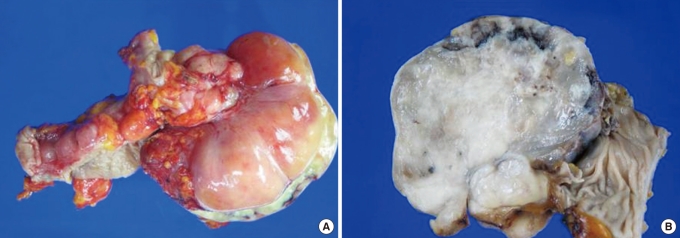
A laparoscopic tumorectomy and segmental resection of the descending colon were performed. Examination of the gross specimen revealed a firm white mass approximately 10 × 9 cm and a 2 × 2 cm mass invading the descending colon (Fig. 5).
The final histopathologic diagnosis was a leiomyosarcoma. The number of mitoses was between 10 and 19 in each of 10 high-power fields; hence, the histologic grade was high. The microscopic examination (× 20 magnification) of a section stained with hematoxylin and eosin revealed a solid tumor mass, well demarcated by a capsule with a peripheral zone. Fascicles of spindle cells with elongated and blunt-ended nuclei were visible at × 100 magnification (Fig. 6). Mitosis was identifiable at a magnification of × 400 (Fig. 6C). Immunohistochemical staining showed positive results for smooth muscle actin and desmin (Fig. 7A, B) and a negative reaction for myosin (Fig. 7C). The patient then received two cycles of mesna, adriamycin, ifosfamide, and dacarbazine (MAID) chemotherapy at the oncology department.
DISCUSSION
Leiomyosarcomas are cancers of the smooth muscle cells, which can arise at any location. The uterus, the retroperitoneum, and the intra-abdominal cavity are common primary sites [3]. This tumor is most commonly seen in the fifth and the sixth decades of life, and the male-to-female ratio is 1:2 [4]. Leiomyosarcomas are characterized by a rapid growth rate and frequent local and systemic recurrences. High-grade leiomyosarcomas frequently metastasize, and the most commonly affected organ is the lung. Prognosis is poor, and the five-year survival rate is 29-36% [5]. In most reported cases, presentations are nonspecific and the results of physical examinations are normal. Radiologic features are nonspecific; therefore, it is difficult to diagnose this cancer preoperatively. A confirmed diagnosis is usually made only postoperatively, which results in delayed diagnosis. Primary renal leiomyosarcomas are sometimes misdiagnosed as renal cell carcinomas [6].
Four years earlier, our patient had also been diagnosed preoperatively as having a renal cell carcinoma based on radiologic examination, the physical examination findings having been nonspecific. The retroperitoneal recurrence of the cancer manifested only as mild diffuse abdominal discomfort. Leiomyosarcomas are distinguished from leiomyomas by cellular pleomorphism, an increased mitotic rate, and the presence of cellular necrosis. Furthermore, in this case, our pathologist conducted immunohistochemical staining, which is useful in the diagnosis of leiomyosarcoma. Immunohistochemical staining revealed positive findings for vimentin and smooth muscle actin and desmin. If a leiomyosarcoma specimen is found to have a pleomorphic component on immunohistochemical staining, it is difficult to distinguish it from a malignant fibrous histiocytoma. However, such heterologous differentiation was not observed in this case [6, 7].
The prognosis for a leiomyosarcoma is usually poor. A good prognosis can be expected only with complete surgical excision (the treatment of choice) [8-10]. Irradiation therapy and chemotherapy do not alter the course of the disease and are not successful in metastatic lesions [9]. A tumor size of less than 5 cm, low histological grade, absence of lymph node metastasis, and radical excision of the mass are all associated with an improved prognosis [1]. To have ensured a better prognosis in this particular case, a radical nephrectomy should have been performed as the first operation. Unfortunately, as the patient had been thought to have a renal cell carcinoma in the preoperative state, she instead underwent a simple left nephrectomy.
Our case is notable and unusual because of the recurrence site and its features. The submucosal-like lesion was detected in the descending colon by using colonoscopy. A submucosal tumor is defined as an elevated or polypoid mass covered with normal mucosa. Several colorectal diseases, such as lipoma, leiomyosarcoma, lymphangioma, and carcinoid tumor, can be detected as lesions mimicking submucosal tumors. CT can help rule out extrinsic compression by abdominal masses. We also confirmed the lesion in the descending colon as being caused by extrinsic compression by performing a CT scan.
As observed in this case, recurrence of a leiomyosarcoma in the retroperitoneum needs to be considered in the differential diagnosis of submucosal tumors in the colon. There is no reported case of recurrence of a renal leiomyosarcoma in the colon. Thus, we report this unusual, but interesting, case of recurrence of a rare renal leiomyosarcoma.




 PDF Links
 PubReader
 XML Download
 Full text via DOI
 Full text via PMC